

Simultaneous variation of the immunodominant outer membrane proteins, MSP2 and MSP3, during anaplasma marginale persistence in vivo
- PMID: 14573687
- PMCID: PMC219554
- DOI: 10.1128/IAI.71.11.6627-6632.2003
Simultaneous variation of the immunodominant outer membrane proteins, MSP2 and MSP3, during anaplasma marginale persistence in vivo
Abstract
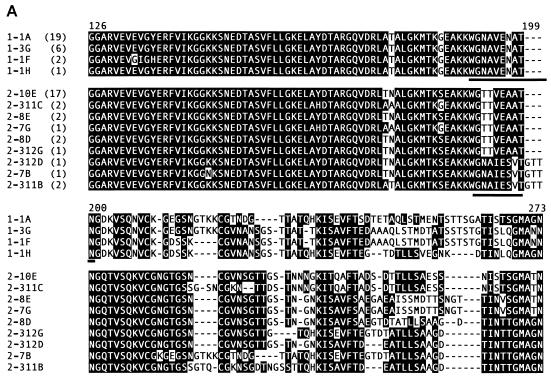
Vector-borne bacterial pathogens persist in the mammalian host by varying surface antigens to evade the existing immune response. To test whether the model of surface coat switching and immune evasion can be extended to a vector-borne bacterial pathogen with multiple immunodominant surface proteins, we examined Anaplasma marginale, a rickettsia with two highly immunogenic outer membrane proteins, major surface protein 2 (MSP2) and MSP3. The simultaneous clearance of variants of the two most immunodominant surface proteins of A. marginale followed by emergence of unique variants indicates that the switch rates and immune selection for MSP2 and MSP3 are sufficiently similar to explain the cyclic bacteremia observed during infection in the immunocompetent host.
Figures


Similar articles
-
Primary Structural Variation in Anaplasma marginale Msp2 Efficiently Generates Immune Escape Variants.Infect Immun. 2015 Nov;83(11):4178-84. doi: 10.1128/IAI.00851-15. Epub 2015 Aug 10. Infect Immun. 2015. PMID: 26259814 Free PMC article.
-
CD4+ T lymphocytes from Anaplasma marginale major surface protein 2 (MSP2) vaccinees recognize naturally processed epitopes conserved in MSP3.Infect Immun. 2004 Jun;72(6):3688-92. doi: 10.1128/IAI.72.6.3688-3692.2004. Infect Immun. 2004. PMID: 15155686 Free PMC article.
-
Emergence of Anaplasma marginale antigenic variants during persistent rickettsemia.Infect Immun. 1999 Nov;67(11):5834-40. doi: 10.1128/IAI.67.11.5834-5840.1999. Infect Immun. 1999. PMID: 10531237 Free PMC article.
-
Antigenic variation in the persistence and transmission of the ehrlichia Anaplasma marginale.Microbes Infect. 2000 Feb;2(2):167-76. doi: 10.1016/s1286-4579(00)00271-9. Microbes Infect. 2000. PMID: 10742689 Review.
-
Antigenic variation and transmission fitness as drivers of bacterial strain structure.Cell Microbiol. 2013 Dec;15(12):1969-75. doi: 10.1111/cmi.12182. Epub 2013 Aug 28. Cell Microbiol. 2013. PMID: 23941262 Free PMC article. Review.
Cited by
-
Differential clearance and immune responses to tick cell-derived versus macrophage culture-derived Ehrlichia chaffeensis in mice.Infect Immun. 2007 Jan;75(1):135-45. doi: 10.1128/IAI.01127-06. Epub 2006 Oct 23. Infect Immun. 2007. PMID: 17060466 Free PMC article.
-
Antigenic Variation in Bacterial Pathogens.Microbiol Spectr. 2016 Feb;4(1):10.1128/microbiolspec.VMBF-0005-2015. doi: 10.1128/microbiolspec.VMBF-0005-2015. Microbiol Spectr. 2016. PMID: 26999387 Free PMC article.
-
Identification of Anaplasma marginale outer membrane protein antigens conserved between A. marginale sensu stricto strains and the live A. marginale subsp. centrale vaccine.Infect Immun. 2011 Mar;79(3):1311-8. doi: 10.1128/IAI.01174-10. Epub 2010 Dec 28. Infect Immun. 2011. PMID: 21189322 Free PMC article.
-
Anaplasma marginale type IV secretion system proteins VirB2, VirB7, VirB11, and VirD4 are immunogenic components of a protective bacterial membrane vaccine.Infect Immun. 2010 Mar;78(3):1314-25. doi: 10.1128/IAI.01207-09. Epub 2010 Jan 11. Infect Immun. 2010. PMID: 20065028 Free PMC article.
-
Antigen variability in Anaplasma phagocytophilum during chronic infection of a reservoir host.Microbiology (Reading). 2012 Oct;158(Pt 10):2632-2641. doi: 10.1099/mic.0.059808-0. Epub 2012 Aug 2. Microbiology (Reading). 2012. PMID: 22859615 Free PMC article.
References
Publication types
MeSH terms
Substances
Grants and funding
LinkOut - more resources
Full Text Sources
Other Literature Sources
