. 2003 Nov;12(11):2476-81.
doi: 10.1110/ps.03267503.

Self-assembly of single integral membrane proteins into soluble nanoscale phospholipid bilayers

Affiliations

Self-assembly of single integral membrane proteins into soluble nanoscale phospholipid bilayers

Timothy H Bayburt et al. Protein Sci. 2003 Nov.

Abstract

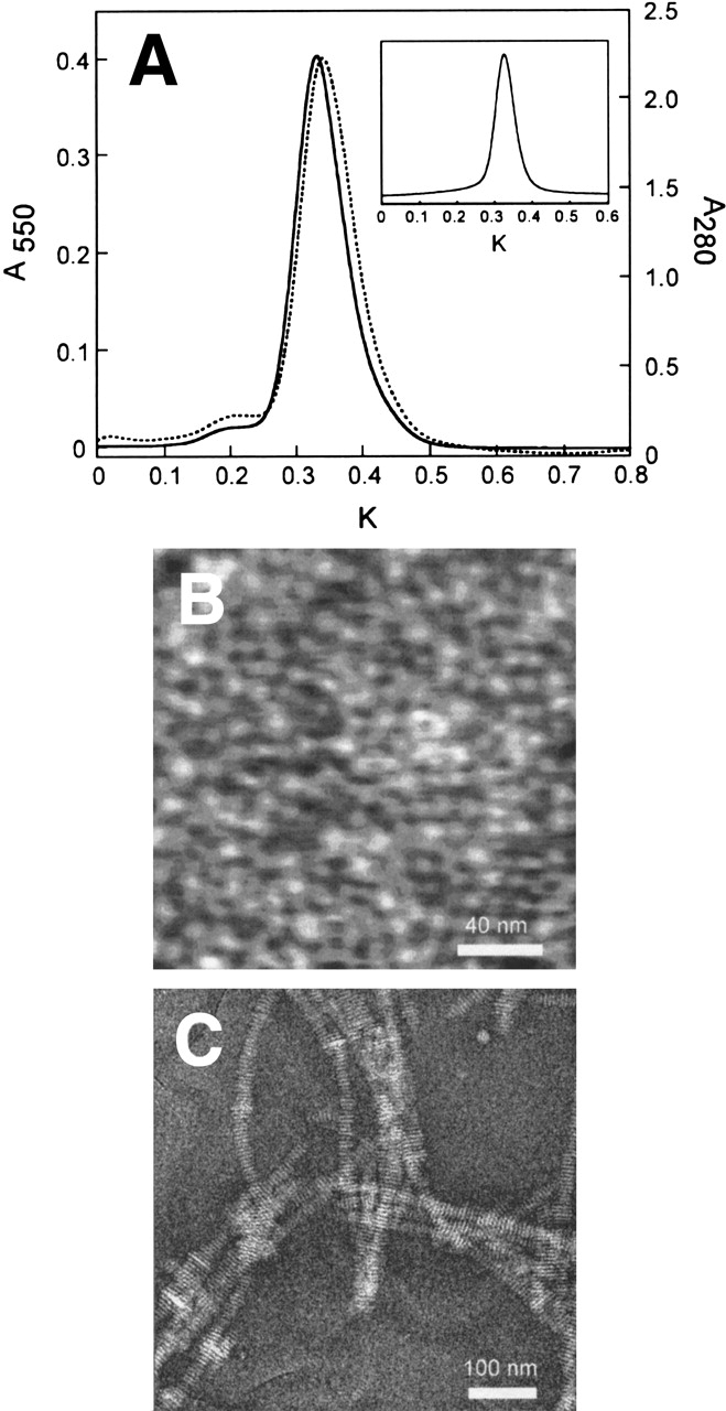
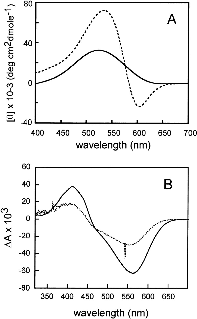
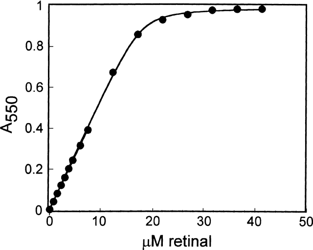
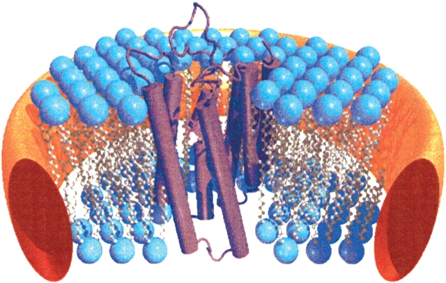
One of the biggest challenges in pharmaceutical research is obtaining integral membrane proteins in a functional, solubilized, and monodisperse state that provides a native-like environment that maintains the spectrum of in vivo activities. Many of these integral membrane proteins are receptors, enzymes, or other macromolecular assemblies that are important drug targets. An example is the general class of proteins composed of seven-transmembrane segments (7-TM) as exemplified by the G-protein-coupled receptors. In this article, we describe a simple system for self-assembling bacteriorhodopsin, as a model protein containing 7-TM helices, with phospholipids to form a nanometer-scale soluble bilayer structure encircled by a 200 amino acid scaffold protein. The result is the single molecule incorporation of an integral membrane protein target into a soluble and monodisperse structure that allows the structural and functional tools of solution biochemistry to be applied.


Figures

Figure 1.
Figure 1.
Gel filtration chromatography of bR Nanodiscs after self-assembly. (A) The chromatogram of the MSP disk formation mixture after detergent removal shows absorbance at 550 nm (solid line) and 280 nm (dotted line). K indicates the partition factor (Velution − Vvoid) / Vinternal. The inset shows reinjection of MSP self-assembled Nanodiscs with monitoring the absorbance at 550 nm. (B) An AFM image of purified bR Nanodiscs on mica. (C) TEM micrograph of phosphotungstate-stained bR Nanodiscs.
Figure 2.
Figure 2.
Optical spectroscopy of bR in Nanodiscs. (A) CD spectra were obtained for disks formed with MSP (solid line) and, for reference, native purple membrane (broken line). (B) Difference spectrum (steady-state illumination minus dark) of bR Nanodiscs and bR proteoliposomes. The absorbance of the bR liposome sample (dashed line) has been normalized to the concentration of bR in the disk sample (solid line, 9.5 μM).
Figure 3.
Figure 3.
Titration of bacteriopsin disks with all-trans-retinal. The initial linear portion of the curve was used to obtain the extinction coefficient at 550 nm. The curve is a fit to a quadratic binding equation, yielding a dissociation constant of 0.3 μM.
Figure 4.
Figure 4.
Fluorescence-detected linear dichroism of probes in surface-oriented assemblies as a function of angle of polarization. Circles indicate DMPC bilayer; squares, DMPC disks; and diamonds, bR DMPC disks. Closed symbols indicate DiIC16(3); open symbols, BODIPY 558/568 C12. Fits of the probe transition dipole polar angle (θ) for bilayer, disk, and bR disk are 74°, 76°, and 76° (DiIC16) and 49, 51° and 50° (BODIPY), respectively.
Figure 5.
Figure 5.
Model of the organization of a Nanodisc. Cartoon shows a cutaway view of a 10-nm-diameter Nanodisc composed of phospholipid (blue) and MSP (orange) containing a molecule of bR (purple). bR was constructed by using coordinates obtained from the Protein Data Bank (PDB ID 1C3W; Luecke et al. 1999; Berman et al. 2000).

Similar articles

Cited by

References

    1. Atkinson, D., Smith, H.M., Dickson, J., and Austin, J.P. 1976. Interaction of apoprotein from porcine high-density lipoprotein with dimyristoyl lecithin, 1: The structure of the complexes. Eur. J. Biochem. 64 541–547. - PubMed
    1. Axelrod, D. 1979. Carbocyanine dye orientation in red cell membrane studied by microscopic fluorescence polarization. Biophys J. 26 557–573. - PMC - PubMed
    1. Bayburt, T.H., Grinkova, Y.V., and Sligar, S.G. 2002. Self-assembly of discoidal phospholipid bilayer nanoparticles with membrane scaffold proteins. Nano Lett. 2 853–856.
    1. Berman, H.M., Westbrook, J., Feng, Z., Gilliland, G., Bhat, T.N., Weissig, H., Shindyalov, I.N., and Bourne, P.E. 2000. The Protein Data Bank. Nucleic Acids Res. 28 235–242. - PMC - PubMed
    1. Brouillette, C.G., Jones, J.L., Ng, T.C., Kercret, H., Chung, B.H., and Segrest, J.P. 1984. Structural studies of apolipoprotein A-I/phosphatidylcholine recombinants by high-field proton NMR, nondenaturing gradient gel electrophoresis, and electron microscopy. Biochemistry 23 359–367. - PubMed

Publication types

LinkOut - more resources