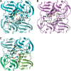
Crystal structure of truncated human betaB1-crystallin
- PMID: 14573871
- PMCID: PMC2366963
- DOI: 10.1110/ps.03265903
Crystal structure of truncated human betaB1-crystallin
Abstract
Crystallins are long-lived proteins packed inside eye lens fiber cells that are essential in maintaining the transparency and refractive power of the eye lens. Members of the two-domain betagamma-crystallin family assemble into an array of oligomer sizes, forming intricate higher-order networks in the lens cell. Here we describe the 1.4 angstroms resolution crystal structure of a truncated version of human betaB1 that resembles an in vivo age-related truncation. The structure shows that unlike its close homolog, betaB2-crystallin, the homodimer is not domain swapped, but its domains are paired intramolecularly, as in more distantly related monomeric gamma-crystallins. However, the four-domain dimer resembles one half of the crystallographic bovine betaB2 tetramer and is similar to the engineered circular permuted rat betaB2. The crystal structure shows that the truncated betaB1 dimer is extremely well suited to form higher-order lattice interactions using its hydrophobic surface patches, linker regions, and sequence extensions.
Figures


Similar articles
-
The N-terminal domain of betaB2-crystallin resembles the putative ancestral homodimer.J Mol Biol. 2000 Dec 1;304(3):253-7. doi: 10.1006/jmbi.2000.4197. J Mol Biol. 2000. PMID: 11090271
-
Human βB2-Crystallin Forms a Face-en-Face Dimer in Solution: An Integrated NMR and SAXS Study.Structure. 2017 Mar 7;25(3):496-505. doi: 10.1016/j.str.2017.02.001. Epub 2017 Feb 23. Structure. 2017. PMID: 28238532 Free PMC article.
-
Circular permutation of betaB2-crystallin changes the hierarchy of domain assembly.Protein Sci. 1998 Jun;7(6):1280-5. doi: 10.1002/pro.5560070602. Protein Sci. 1998. PMID: 9655330 Free PMC article.
-
Probing the structure and interactions of crystallin proteins by NMR spectroscopy.Prog Retin Eye Res. 1999 Jul;18(4):431-62. doi: 10.1016/s1350-9462(98)00027-5. Prog Retin Eye Res. 1999. PMID: 10217479 Review.
-
Chemical Properties Determine Solubility and Stability in βγ-Crystallins of the Eye Lens.Chembiochem. 2021 Apr 16;22(8):1329-1346. doi: 10.1002/cbic.202000739. Epub 2021 Feb 10. Chembiochem. 2021. PMID: 33569867 Free PMC article. Review.
Cited by
-
Cataract-causing mutation S228P promotes βB1-crystallin aggregation and degradation by separating two interacting loops in C-terminal domain.Protein Cell. 2016 Jul;7(7):501-15. doi: 10.1007/s13238-016-0284-3. Epub 2016 Jun 18. Protein Cell. 2016. PMID: 27318838 Free PMC article.
-
CRYBA4, a novel human cataract gene, is also involved in microphthalmia.Am J Hum Genet. 2006 Oct;79(4):702-9. doi: 10.1086/507712. Epub 2006 Aug 17. Am J Hum Genet. 2006. PMID: 16960806 Free PMC article.
-
Eye lens β-crystallins are predicted by native ion mobility-mass spectrometry and computations to form compact higher-ordered heterooligomers.Structure. 2023 Sep 7;31(9):1052-1064.e3. doi: 10.1016/j.str.2023.06.013. Epub 2023 Jul 14. Structure. 2023. PMID: 37453416 Free PMC article.
-
Ubiquitin proteasome pathway-mediated degradation of proteins: effects due to site-specific substrate deamidation.Invest Ophthalmol Vis Sci. 2010 Aug;51(8):4164-73. doi: 10.1167/iovs.09-4087. Epub 2010 Jun 30. Invest Ophthalmol Vis Sci. 2010. PMID: 20592226 Free PMC article.
-
Molecular and structural analysis of genetic variations in congenital cataract.Mol Vis. 2013 Nov 24;19:2436-50. eCollection 2013. Mol Vis. 2013. PMID: 24319337 Free PMC article.
References
-
- Ajaz, M.S., Ma, Z., Smith, D.L., and Smith, J.B. 1997. Size of human lens β-crystallin aggregates are distinguished by N-terminal truncation of βB1. J. Biol. Chem. 272 11250–11255. - PubMed
-
- Bateman, O.A., Lubsen, N.H., and Slingsby, C. 2001. Association behaviour of human βB1-crystallin and its truncated forms. Exp. Eye Res. 73 321–331. - PubMed
-
- Bateman, O.A., Sarra, R., Slingsby, C., van Genesen, S.T., Kappé, G., and Lubsen, N.H. 2003. The stability of human acidic β-crystallin oligomers and hetero-oligomers. Exp. Eye Res. (in press). - PubMed
-
- Bax, B., Lapatto, R., Nalini, V., Driessen, H., Lindley, P.F., Mahadevan, D., Blundell, T.L., and Slingsby, C. 1990. X-ray analysis of βB2-crystallin and evolution of oligomeric lens proteins. Nature 347 776–780. - PubMed
-
- Berbers, G.A.M., Boermann, O.C., Bloemendal, H., and de Jong, W.W. 1982. Primary gene products of bovine β-crystallin and reassociation behaviour of its aggregates. Eur. J. Biochem. 128 495–502. - PubMed
Publication types
MeSH terms
Substances
Associated data
- Actions
- Actions
LinkOut - more resources
Full Text Sources
Molecular Biology Databases

