. 2004 Feb;53(2):118-24.
doi: 10.1007/s00262-003-0437-0. Epub 2003 Oct 22.

In vitro biological activities of transmembrane superantigen staphylococcal enterotoxin A fusion protein

Affiliations

In vitro biological activities of transmembrane superantigen staphylococcal enterotoxin A fusion protein

Wenxue Ma et al. Cancer Immunol Immunother. 2004 Feb.

Abstract

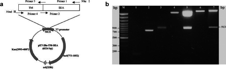
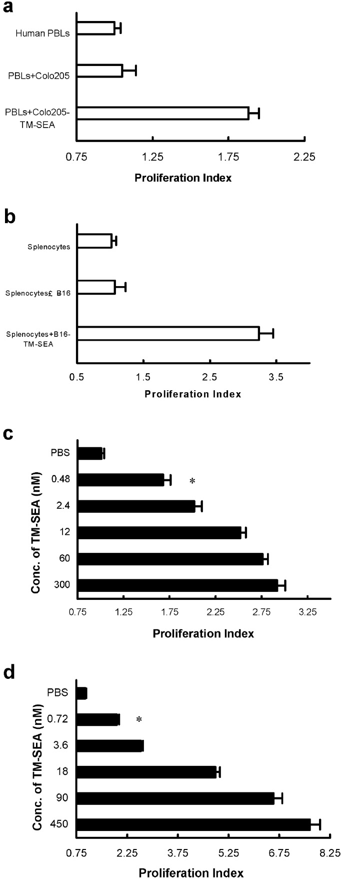
The bacterial superantigen staphylococcal enterotoxin A (SEA) stimulates T cells bearing certain TCR Vbeta domains when binding to MHC II molecules, and is a potent inducer of CTL activity and cytokine production. Antibody-targeted SEA such as C215 Fab-SEA and C242 Fab-SEA has been investigated for cancer therapy in recent years. We have previously reported significant tumor inhibition and prolonged survival time in tumor-bearing mice treated with a combination of both C215Fab-SEA and Ad IL-18 (Wang et al., Gene Therapy 8:542-550, 2001). In order to develop SEA as an universal biological preparation in cancer therapy, we first cloned a SEA gene from S. aureus (ATCC 13565) and a transmembrane (TM) sequence from a c- erb-b2 gene derived from human ovarian cancer cell line HO-8910, then generated a TM-SEA fusion gene by using the splice overlap extension method, and constructed the recombinant expression vector pET-28a-TM-SEA. Fusion protein TM-SEA was expressed in E. coli BL21(DE3)pLysS and purified by using the histidine tag in this vector. Purified TM-SEA spontaneously associated with cell membranes as detected by flow cytometry. TM-SEA stimulated the proliferation of both human PBLs and splenocytes derived from C57BL/6 (H-2b) mice in vitro. This study thus demonstrated a novel strategy for anchoring superantigen SEA onto the surfaces of tumor cells without any genetic manipulation.


Figures

Fig. 1
Fig. 1
a Schematic representation of the expression vector containing TM-SEA. Arrows indicate relative positions of the primers. Figure not drawn to scale. TM-SEA was generated by splice overlap extension PCR, subcloning of TM-SEA fusion gene into pET-28a vector results in a N-terminal histidine sequence. Kan denotes the kanamycin resistance gene, ori origin of DNA replication. b Electrophoresis of PCR product of TM, SEA, TM-SEA gene and analysis of the recombinant plasmid digested with Nhe I and Hind III. TM fragment, SEA gene, and TM-SEA fusion gene were amplified by PCR, respectively; the recombinant pGEM-T and pET-28a plasmids were digested with Nhe I and Hind III, all the PCR and digested products were analyzed by electrophoresis in a 1.7% agarose gel containing 0.5 μg/ml ethidium bromide. M 100 bp DNA ladder plus, Lane 1 TM PCR product (155 bp), lane 2 SEA PCR product (796 bp), lane 3 TM-SEA PCR product (922 bp), lane 4 empty pGEM-T vector, lane 5 recombinant pGEM-T-TM-SEA vector digested with Nhe I and Hind III (913 bp), lane 6 empty pET-28a vector, lane 7 recombinant pET-28a-TM-SEA vector digested with Nhe I and Hind III (913 bp)
Fig. 2
Fig. 2
a Western blotting analysis of the target protein TM-SEA from BL21(DE3)pLysS-pET-28a-TM-SEA strain. PVDF membrane was immunoblotted with antisera raised against the following samples order: Lane 1 purified SEA (Toxin Technology, USA) 30 ng/well as a positive control, lane 2 BL21(DE3)pLysS-pET-28a vector, lane 3 engineering strain BL21(DE3)pLysS-pET-28a-TM-SEA without inducement of IPTG, lanes 4–7 engineering strain BL21(DE3)pLysS-pET-28a-TM-SEA was induced 1 h, 3 h, 5 h, and 7 h, respectively, by IPTG with the final concentration of 1.0 mmol. b Western blotting analysis of the purified TM-SEA fusion protein. The samples were resolved on a 12% polyacrylamide gel in the following order: Lane 1 purified SEA (30 ng loaded) as a positive control, lane 2 supernatant of the lysates of engineering strain BL21(DE3)pLysS-pET-28a-TM-SEA containing the target protein, lane 3 unbound protein, lane 4 purified target TM-SEA protein
Fig. 3a–c
Fig. 3a–c
TM-SEA spontaneously anchored onto the tumor cells. B16 cells were incubated with TM-SEA fusion protein for 4 h at 37°C. After washing twice, the cells were incubated with first antibody (rabbit anti-SEA IgG) for 1 hr at 37°C, and then incubated with the FITC-labeled second antibody (antirabbit IgG/FITC) for 40 min. The cells were harvested and detected by flow cytometry. B16 cells incubated with SEA protein (a) and TM-SEA protein (b) for 4 h, then incubated with the antibodies respectively. The cells were washed and used immediately. Of the B16 cells, 94.9% were positive to the TM protein. For detecting the stability of TM-SEA fusion protein anchored onto the cell membrane (c), B16 cells were incubated with TM-SEA fusion protein for 4 h, the cells were washed and incubated with medium for 4 h, then treated with the antibodies respectively, 87% of B16 cells were positive to the TM-SEA protein
Fig. 4a, b
Fig. 4a, b
Proliferative effects of the purified TM-SEA protein on lymphocytes in vitro. Human PBLs (a) and splenocytes derived from C57BL/6 mice (b) were cultured (with a final concentration of 5×10⁶ cells/ml, 0.1 ml for each well) in flat-bottomed 96-well plates for 48 h in the presence of TM-SEA and/or SEA with 10-fold serial concentration. After culturing for 48 h, the proliferation of lymphocytes was determined by MTT assay. PHA and Con A were as the positive control respectively, and PBS was as the negative controls. Value on the y-axis represents the average proliferation index (PI), error bars show SD of triplicate cultures; *p<0.01 vs incubated with PBS
Fig. 5a–d
Fig. 5a–d
TM-SEA stimulates lymphocyte proliferation. Either inactivated Colo205 cells or B16 cells incubated with TM-SEA of 0.3 μmol or 0.45 μmol, respectively, for 48 h, stimulate the proliferation of human PBLs (a) or splenocytes derived from C57BL/6 mice (b); *p<0.01 vs the controls, q tests. c Tumor cells incubated with TM-SEA stimulate the lymphocytes proliferation: Colo205 cells inactivated with MMC were preincubated with fivefold serial dilutions of TM-SEA, and added at a 1:1 Colo205 to PBLs (T:L) ratio to 2×10⁵ PBLs/well in a 96-well plate. d B16 cells inactivated with MMC were preincubated with fivefold serial dilutions of TM-SEA, and added at a 1:1 B16 to splenocytes (T:L) ratio to 2×10⁵ splenocytes/well in a 96-well plate. Values on the x-axis represent the proliferation index ± SD of triplicate cultures; *p<0.01 vs the controls, q tests

References

    1. Johnson FASEB J. 1991;5:2706. - PubMed
    1. Betley J Bacteriology. 1988;170:34. - PMC - PubMed
    1. Fraser Mol Med Today. 2000;6:125. doi: 10.1016/S1357-4310(99)01657-3. - DOI - PubMed
    1. Hudson J Exp Med. 1993;177:175. - PMC - PubMed
    1. Klarnet J Exp Med. 1989;169:457. - PMC - PubMed

Publication types

MeSH terms

LinkOut - more resources