. 2003 Nov;73(5):1170-7.
doi: 10.1086/379085. Epub 2003 Oct 21.

Protein-truncating mutations in ASPM cause variable reduction in brain size

Affiliations

Protein-truncating mutations in ASPM cause variable reduction in brain size

Jacquelyn Bond et al. Am J Hum Genet. 2003 Nov.

Abstract

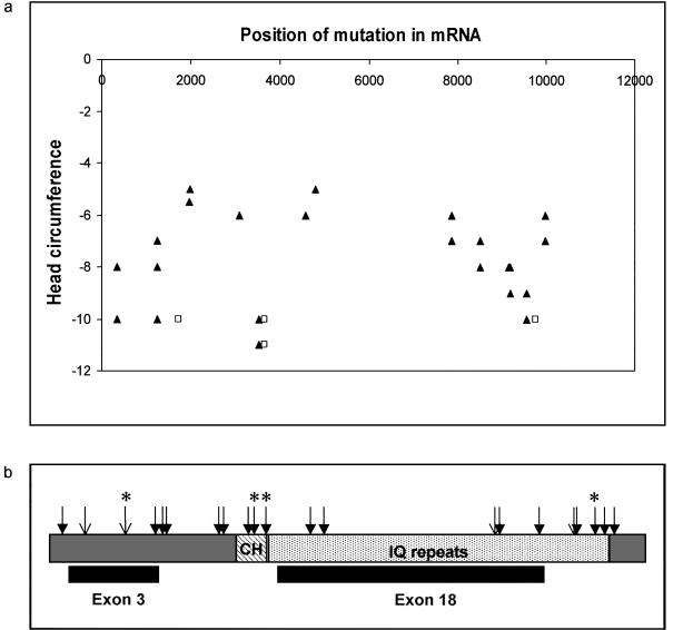
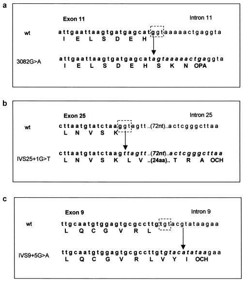
Mutations in the ASPM gene at the MCPH5 locus are expected to be the most common cause of human autosomal recessive primary microcephaly (MCPH), a condition in which there is a failure of normal fetal brain development, resulting in congenital microcephaly and mental retardation. We have performed the first comprehensive mutation screen of the 10.4-kb ASPM gene, identifying all 19 mutations in a cohort of 23 consanguineous families. Mutations occurred throughout the ASPM gene and were all predicted to be protein truncating. Phenotypic variation in the 51 affected individuals occurred in the degree of microcephaly (5-11 SDs below normal) and of mental retardation (mild to severe) but appeared independent of mutation position.


Figures

Figure  1
Figure 1
A northern Pakistani individual with MCPH. This individual has the sloping forehead typical of those with MCPH. He and other affected members of his family have an ASPM mutation described in this report.
Figure  2
Figure 2
The position of ASPM mutations identified in families with MCPH5. a, ASPM mutation position compared to the degree of microcephaly. On the x axis, the position of the ASPM gene mutations (listed in table C1) is shown, with the gene sequence aligned 5′→3′. The head circumference of individuals with MCPH5 in whom a mutation was found in this study is shown on the y axis. The head circumference is shown as a SD from the mean (corrected for age and sex). Some points represent more than one affected individual. Those individuals who were judged to exhibit severe mental retardation are shown as unfilled squares, whereas those with mild or moderate mental retardation are represented as filled triangles. There is no relationship between mutation position and head circumference or the degree of mental retardation. b, The ASPM protein as a schematic with the major protein domains depicted; CH (diagonally striped box) depicts the calponin homology domain, and the IQ domain repeats (light gray hatched box) are represented. The position of the large nonterminal exons 3 and 18 are indicated beneath the corresponding region they encode. Novel mutations identified in this study are shown as filled arrows. Unfilled arrows indicate mutations reported in the work of Bond et al. (2002). Mutations found in more than one family are denoted with an asterisk.
Figure  3
Figure 3
Mutations in ASPM splice-donor motifs. The splice-donor site (the last guanine of the exon in boldface and the guanine-thymidine of the intron in plain text) is shown as a broken boxed area drawn onto the wild-type genomic sequence. In each panel, the upper genomic sequence is wild type, and the lower contains the mutation, indicated by an arrow. Bases that are exonic are shown in boldface, and those that are potentially translated intronic sequence are shown in italicized boldface. The normal and novel amino acids introduced by the failure to splice and the translation read-through are indicated beneath the wild-type and mutation sequences, respectively. The predicted premature stop codons caused by the mutations are indicated as OPA (opal) and OCH (ochre). a, The exon 11 splice-donor site is removed by a G→A mutation at base 3082, thus allowing the incorporation of an additional three amino acids into the protein before translation termination. b, The point mutation G→T at IVS25 +1 eliminates the splice-donor site, allowing translation to continue until a stop signal is reached 29 codons downstream. c, The point mutation G→A at IVS9+5 is predicted to remove the splice-donor site, therefore translation continues for a further two amino acids until a translation-termination motif is reached.

References

Electronic-Database Information

    1. Berkeley Drosophila Genome Project, http://www.fruitfly.org/seq_tools/splice.html (for splice prediction)
    1. INFOBIOGEN, http://www.infobiogen.fr/services/analyseq/cgi-bin/traduc_in.pl (for translation of nucleotide sequence to amino acids)
    1. National Center for Biotechnology Information (NCBI), Entrez search engine, http://www.ncbi.nlm.nih.gov/entrez/query.fcgi?db=Nucleotide (for ASPM)
    1. Online Mendelian Inheritance in Man (OMIM), http://www.ncbi.nlm.nih.gov/omim/ (for MCPH [MIM 251200] and ASPM [MIM 650481])

References

    1. Aicardi J (1998) Malformations of the central nervous system. In: Diseases of the nervous system in childhood, 2nd ed. Mac Keith, London, pp 90–91
    1. Bond J, Roberts E, Mochida GH, Hampshire DJ, Scott S, Askham JM, Springell K, Mahadevan M, Crow YJ, Markham AF, Walsh CA, Woods CG (2002) ASPM is a major determinant of cerebral cortical size. Nat Genet 32:316–320 - PubMed
    1. Bundey S (1997) Abnormal Mental Development. In: Rimoin DL, Connor JM, Pyeritz RE (eds) Emery and Rimoin’s principles and practice of medical genetics, 3rd ed. Churchill Livingstone, New York, pp 730–731
    1. Craig R, Norbury C (1998) The novel murine calmodulin-binding protein Sha1 disrupts mitotic spindle and replication checkpoint functions in fission yeast. J Cell Sci 111:3609–3619 - PubMed
    1. Gonzalez C, Molina I, Casal J, Ripoll P (1989) Gross genetic dissection and interaction of the chromosomal region 95E;96F of Drosophila melanogaster. Genetics 123:371–377 - PMC - PubMed

MeSH terms

Associated data