. 2004 Feb 15;103(4):1342-7.
doi: 10.1182/blood-2003-09-3051. Epub 2003 Oct 23.

Role of tissue factor and protease-activated receptors in a mouse model of endotoxemia

Affiliations

Role of tissue factor and protease-activated receptors in a mouse model of endotoxemia

Rafal Pawlinski et al. Blood. .

Abstract

Sepsis is associated with a systemic activation of coagulation and an excessive inflammatory response. Anticoagulants have been shown to inhibit both coagulation and inflammation in sepsis. In this study, we used both genetic and pharmacologic approaches to analyze the role of tissue factor and protease-activated receptors in coagulation and inflammation in a mouse endotoxemia model. We used mice expressing low levels of the procoagulant molecule, tissue factor (TF), to analyze the effects of TF deficiency either in all tissues or selectively in hematopoietic cells. Low TF mice had reduced coagulation, inflammation, and mortality compared with control mice. Similarly, a deficiency of TF expression by hematopoietic cells reduced lipopolysaccharide (LPS)-induced coagulation, inflammation, and mortality. Inhibition of the down-stream coagulation protease, thrombin, reduced fibrin deposition and prolonged survival without affecting inflammation. Deficiency of either protease activated receptor-1 (PAR-1) or protease activated receptor-2 (PAR-2) alone did not affect inflammation or survival. However, a combination of thrombin inhibition and PAR-2 deficiency reduced inflammation and mortality. These data demonstrate that hematopoietic cells are the major pathologic site of TF expression during endotoxemia and suggest that multiple protease-activated receptors mediate crosstalk between coagulation and inflammation.


Figures

Figure 1
Figure 1. Role of TF in LPS-induced lethality
(A) TAT complex levels were measured at 6 hours in mTF+/−/hTF+ and low TF mice injected with either saline or LPS (10 mg/kg). Data are shown as mean ± SE (n = 5 per group). (B) Kaplan-Meier plot of the survival of mTF+/−/hTF+ mice (solid line; n = 22) and low TF mice (dashed line; n = 21) after administration of LPS (10 mg/kg). (C–D) □ indicates TNF-α and IL-6 levels in mTF+/−/hTF+ mice, and ■ indicates levels in low TF mice (mean ± SE; n = at least 4 per group).
Figure 2
Figure 2. Role of hematopoietic cell TF expression in LPS-induced lethality
WT mice were irradiated and reconstituted with bone marrow from either low TF mice or littermate mTF+/−/hTF+ control mice. (A) PCR analysis of DNA from peripheral blood cells from WT mice that underwent transplantation with bone marrow from either mTF+/−/hTF+ or low TF mice was used to demonstrate reconstitution of the donor mice with recipient bone marrow 6 weeks after the transplantation. PCR was performed for the WT TF (WT) allele. Glyceraldehyde-3-phosphate dehydrogenase (GAPDH) was used as a loading control. First lanes contain molecular weight standards. (B) LPS-induced TAT complex levels (6 hours) (mean ± SE; n = 4 to 5 per group). (C) Survival of endotoxemic mice (5 mg/kg LPS intraperitoneally) reconstituted with bone marrow from either mTF+/−/hTF+ mice (solid line; n = 20) or low TF mice (dashed line; n = 22). For survival studies, we used mice that underwent transplantation with bone marrow derived from 3 independent donors for each genotype. (D) LPS-induced IL-6 expression in WT mice receiving bone marrow from mTF+/−/hTF+ (□) or low TF (■) mice (mean ± SE; n = 4 per group). Differences between the 2 groups were not statistically significant.
Figure 3
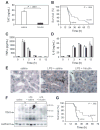
Figure 3. Effect of administration of hirudin and ancrod on endotoxemic mice
(A) TAT complex levels were measured at 6 hours in endotoxemic mice treated with saline or hirudin. Data are shown as mean ± SE (more than 4 mice per group). (B) Survival of WT mice injected with LPS and treated with either saline (solid line; n = 15) or hirudin (dashed line; n = 15). (C–D) TNF-α and IL-6 levels in saline-treated (□) and hirudin-treated (■) endotoxemic mice are shown. Data are shown as mean ± SE (more than 4 mice per group). (E) Immunohistochemical analysis of fibrin deposition in the liver. Mice were injected with saline, LPS and saline, or LPS and hirudin, and livers were collected at 8 hours. Fibrin(ogen) (brown color) was detected using a rabbit antifibrin(ogen) polyclonal antibody. Original magnification, × 400. Representative photomicrographs from 1 of 3 mice per group are shown. (F) Fibrin deposition in the liver. Mice (3 per group) were injected with saline, LPS and saline, or LPS and hirudin. Fibrin was detected in livers at 8 hours by Western blotting using an antifibrin monoclonal antibody. A pancadherin antibody was used to monitor protein loading. (G) Survival of wild-type mice injected with LPS and treated with either saline (solid line; n = 12) or ancrod (dashed line; n = 12). P values are shown.
Figure 4
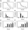
Figure 4. Role of PARs in LPS-induced lethality
(A) Survival of endotoxemic PAR-2+/+ (solid line; n = 10) and PAR-2−/− (dashed line; n = 9) mice. (B–C) LPS induction of TNF-α and IL-6 expression in PAR-2+/+ (□) and PAR-2−/− (■). Data are shown as mean ± SE (3 to 6 mice per group). (D) Survival of endotoxemic PAR-1+/+ (solid line; n = 11) and PAR-1−/− (dashed line; n = 13). (E–F) LPS induction of TNF-α and IL-6 expression in PAR-1+/+ (□) and PAR-1−/− (■). Data are shown as mean ± SE (3 to 6 mice per group). No statistically significant differences were observed.
Figure 5
Figure 5. Effect of combined hirudin treatment and PAR-2 deficiency
(A) Survival of LPS-treated PAR-2+/+ mice with saline (solid line; n = 10), LPS-treated PAR-2+/+ mice with hirudin (dashed line; n = 17), and LPS-treated PAR-2−/− mice with hirudin (dotted line; n = 17). Hirudin statistically increased the survival time of PAR-2+/+ mice (P =.0001) and PAR-2−/− mice (P %.0001) compared with saline-treated PAR-2+/+ mice. The difference in survival between PAR-2+/+ and PAR-2−/− mice treated with hirudin is also statistically significant (P =.037). (B) LPS-induced IL-6 expression (8 hours) in PAR-2+/+ and PAR-2−/− mice treated with either saline or hirudin. Data are shown as mean ± SE (more than 6 mice per group).
Figure 6
Figure 6. Coagulation proteases increase inflammation during endotoxemia via PARs
FVIIa and FXa activation of PAR-2 and thrombin (IIa) activation of PAR-1 and PAR-4 may enhance inflammation. Thrombin also cleaves fibrinogen to fibrin and activates platelets by cleavage of PAR-3/4. Inhibitors of the TF-FVIIa complex are shown: α F Ab (anti-TF antibody), FVIIai (active site-inhibited FVIIa), and TFPI-1. Hirudin inhibits thrombin, and ancrod depletes fibrinogen.

Similar articles

Cited by

References

    1. Parrillo JE. Pathogenetic mechanisms of septic shock. N Engl J Med. 1993;328:1471–1477. - PubMed
    1. Riedemann NC, Guo RF, Ward PA. Novel strategies for the treatment of sepsis. Nat Med. 2003;9:517–524. - PubMed
    1. Creasey AA, Chang AC, Feigen L, Wun TC, Hinshaw LB. Tissue factor pathway inhibitor reduces mortality from Escherichia coli septic shock. J Clin Invest. 1993;91:2850–2856. - PMC - PubMed
    1. Taylor FB, Jr, Emerson TE, Jr, Jordan R, Chang AK, Blick KE. Antithrombin-III prevents the lethal effects of Escherichia coli infusion in baboons. Circ Shock. 1998;26:227–235. - PubMed
    1. Taylor FB, Jr, Chang A, Esmon CT, D’Angelo A, Vigano-D’Angelo S, Blick KE. Protein C prevents the coagulopathic and lethal effects of Escherichia coli infusion in the baboon. J Clin Invest. 1987;79:918–925. - PMC - PubMed

Publication types

MeSH terms