. 2004 Jan 9;279(2):1359-67.
doi: 10.1074/jbc.M306248200. Epub 2003 Oct 23.

Activation of the pro-survival phosphatidylinositol 3-kinase/AKT pathway by transforming growth factor-beta1 in mesenchymal cells is mediated by p38 MAPK-dependent induction of an autocrine growth factor

Affiliations

Activation of the pro-survival phosphatidylinositol 3-kinase/AKT pathway by transforming growth factor-beta1 in mesenchymal cells is mediated by p38 MAPK-dependent induction of an autocrine growth factor

Jeffrey C Horowitz et al. J Biol Chem. .

Abstract

Transforming growth factor-beta1 (TGF-beta1) is a multifunctional cytokine involved in differentiation, growth, and survival of mesenchymal cells while inhibiting growth/survival of most other cell types. The mechanism(s) of pro-survival signaling by TGF-beta1 in mesenchymal cells is unclear. In this report, we demonstrate that TGF-beta1 protects against serum deprivation-induced apoptosis of mesenchymal cells isolated from patients with acute lung injury and of normal human fetal lung fibroblasts (IMR-90). TGF-beta receptor(s)-activated signaling in these cells involves rapid activation of the Smad and p38 MAPK pathways within minutes of TGF-beta1 treatment followed by a more delayed activation of the pro-survival phosphatidylinositol 3-kinase-protein kinase B (PKB)/Akt pathway. Pharmacological inhibition of p38 MAPK with SB203580 or expression of a p38 kinase-deficient mutant protein inhibits TGF-beta1-induced PKB/Akt phosphorylation. Conditioned medium from TGF-beta1-treated cells rapidly induces PKB/Akt activation in an SB203580- and suramin-sensitive manner, suggesting p38 MAPK-dependent production of a secreted growth factor that activates this pro-survival pathway by an autocrine/paracrine mechanism. Inhibition of the phosphatidylinositol 3-kinase-PKB/Akt pathway blocks TGF-beta1-induced resistance to apoptosis. These results demonstrate the activation of a novel TGF-beta1-activated pro-survival/anti-apoptotic signaling pathway in mesenchymal cells/fibroblasts that may explain cell-specific actions of TGF-beta1 and provide mechanistic insights into its pro-fibrotic and tumor-promoting effects.


Figures

Fig. 1
Fig. 1. TGF-β1 protects human lung mesenchymal cells/fibroblasts from serum deprivation-induced apoptosis.
A, mesenchymal cells (ALI-MC) isolated from the lung of a patient with acute lung injury were cultured in 96-well cell culture plates in the presence of 10% FBS and grown to 80% confluence. Cells were then growth-arrested for 48 h and treated with/without TGF-β1 (2 ng/ml) in the presence or absence of serum for an additional 72 h. Apoptosis assays were performed as described under “Materials and Methods.” n = 4 for each group; *, p < 0.001 compared with serum/control; **, p < 0.001 compared with serum-deprived/control. B, normal fetal lung fibroblasts (IMR-90) were cultured, treated with/without TGF-β1, and assayed for apoptosis under identical conditions as described above. n = 4 for each group; *, p < 0.001 compared with serum/control; **, p < 0.01 compared with serum-deprived/control.
Fig. 2
Fig. 2. TGF-β1 induces time-dependent phosphorylation of PKB/Akt in human lung mesenchymal cells/fibroblasts.
A, mesenchymal cells (ALI-MC) isolated from the lung of a patient with acute lung injury were cultured in the presence of 10% FBS, grown to 80% confluence, and then growth-arrested for 48 h. Cells were then treated with TGF-β1 (2 ng/ml) for the indicated times prior to cell lysis. Cell lysates were then subjected to SDS-PAGE and immunoblotted with phospho-specific antibodies to serine-473 of PKB/Akt (S473 phos-pho-Akt). The same blots were stripped and probed for total Akt. B, normal fetal lung fibroblasts (IMR-90) were similarly analyzed for induction of PKB/Akt phosphorylation in response to TGF-β1 stimulation.
Fig. 3
Fig. 3. TGF-β1 induces rapid activation of both Smad2 and p38 MAPK pathways in human lung fibroblasts.
A, cultured human lung fibroblasts (IMR-90) were grown to near-confluence, growth-arrested for 48 h in 0.01% serum, and treated with TGF-β1 (2 ng/ml) for the indicated times prior to cell lysis. Whole cell lysates were subjected to SDS-PAGE and immunoblotted with antibodies to phospho-Smad2 and phospho-p38 MAPK. Each blot was then stripped and probed for total Smad2 or p38 MAPK. B, densitometric analyses of the levels of Smad2 and p38 MAPK phosphorylations represented in A.
Fig. 4
Fig. 4. Pharmacological inhibition of p38 MAP kinase inhibits TGF-β1-induced PKB/Akt phosphorylation in human lung fibro-blasts.
A, mesenchymal cells isolated from the lung of a patient with acute lung injury (ALI-MC) were grown to near-confluence and growth-arrested for 48 h prior to treatment with/without TGF-β1 (2 ng/ml for 12 h) in the presence/absence of the indicated protein kinase inhibitors. Cell lysates were then obtained and subjected to SDS-PAGE followed by immunoblotting against phosphorylated (serine 473) PKB/Akt (S473 phospho-Akt). The blot was stripped and probed for total Akt. B, densitometric analyses of the Western blots in A. C, human lung fibroblasts (IMR-90) were similarly cultured and treated ± TGF-β1 (2 ng/ml) ± protein kinase inhibitors as described above. Western blot analyses for Ser-473-phospho-Akt and total Akt are shown. D, densitometric analyses of the Western blots in C. PD, PD98059 (20 μm); SB, SB203580 (6 μm); Jnk-I, SP600125 (100 nm); Rho-I, Y27632 (15 nm); LY, LY294002 (10 μ m); and WM, wortmannin (50 nm).
Fig. 5
Fig. 5. The p38 MAPK inhibitor, SB203580, blocks TGF-β1-induced phosphorylation of Akt in a dose-dependent manner.
A, cultured IMR-90 cells were grown to near-confluence, growth-arrested for 48 h, and then stimulated with/without TGF-β1 (2 ng/ml) in the presence of the indicated concentrations of the p38 MAPK inhibitor, SB203580. Cell lysates were obtained 12 h after treatment and subjected to SDS-PAGE followed by immunoblotting with an antibody to phospho(serine 473)-specific PKB/Akt. The blot was stripped and probed with antibody to total Akt. B, densitometric analyses of phospho-Akt:total Akt Western blots represented in A.
Fig. 6
Fig. 6. Functional p38 MAPK activity is required for TGF-β1-induced PKB/Akt phosphorylation.
A, IMR-90 cells stably transfected with control plasmid (pcDNA), a kinase-deficient p38 MAPK construct (p38-KM), or wild-type MKK3 (MKK3) were grown to near-confluence, growth-arrested for 48 h prior to treatment with or without TGF-β1 (2 ng/ml), and cell lysates obtained at 1, 12, and 16 h. Phosphorylation of p38 MAPK was assessed at 1 h following TGF-β1 treatment by Western immunoblotting with antibodies to phospho- and total p38 MAPK (top panels). p38 MAP kinase activity was assessed at 1 h following TGF-β1 treatment by in vitro phosphorylation of the p38 MAPK substrate, ATF-2, as described under “Materials and Methods” (middle top panel). Phosphorylation of PKB/Akt was assessed at 12 h following TGF-β1 treatment by Western immunoblotting with antibodies to phospho- and total Akt (middle bottom panels). Cell lysates obtained 16 h after treatment with TGF-β1 were subjected to Western immunoblotting for α-smooth muscle actin (α-SMA), a marker of myofibroblast differentiation (bottom panel). B, densitometric analyses of the phospho-Akt:total Akt blots represented in A. C, stably transfected cells were cultured to 80% confluence, growth-arrested in DMEM with 0.01% FBS for 48 h, treated with/without TGF-β1 (2 ng/ml) for 2 days. Representative photographs demonstrate cellular morphology under these serum-deprived conditions.
Fig. 7
Fig. 7. TGF-β1 induces p38 MAPK-dependent production/secretion of a growth factor that mediates PKB/Akt activation in an autocrine manner.
A, IMR-90 cells were treated with/without TGF-β1, and the conditioned medium was collected at 16 h. Conditioned media were added to a different set of “reporter” IMR-90 cell cultures for the shorter periods indicated. Cell lysates were then subjected to SDS-PAGE and immunoblotted with antibodies against phosphorylated (serine 473) PKB/Akt (S473 phospho-Akt); blots were stripped and re-probed for total Akt. Suramin (300 μ m, Sur) was pre-added to reporter cells 30 min prior to incubation with conditioned medium for 15 min. Control cells (C) that received no conditioned medium are also represented. B, densitometric analyses of the ratio of phospho-Akt to total Akt determined from immunoblots in A. C, conditioned media were collected from IMR-90 cells treated with TGF-β1 in the presence/absence of inhibitors of p38 MAPK (SB203580, 10 μ m) or ERK1/2 MAPK (PD98049, 20 μ m) for 16 h. Conditioned medium was then added to reporter IMR-90 cells that had been serum-deprived for 24 h. Cells were then harvested at 15 min and lysates subjected to Western blot analysis for phosphorylated (serine 473) PKB/Akt (S473 phospho-Akt); blots were stripped and probed for total Akt.
Fig. 8
Fig. 8. TGF-β1-induced protection from serum deprivation-induced apoptosis of lung fibroblasts is dependent on the PI3K/Akt pathway.
A, IMR-90 cells were grown to near-confluence in a 96-well ELISA culture plate and growth-arrested for 48 h in 0.01% serum prior to treatment with TGF-β1 ± LY294002 (10 μ m) as indicated for 5 days. Apoptosis rates were measured using an ELISA for single-stranded DNA as described under “Materials and Methods.” Findings represent one of three independent experiments. Values are mean ± S.E., n = 3. *, p < 0.05 compared with untreated controls. B, cultured IMR-90 cells were grown to 50% confluence, growth-arrested in 0.01% serum for 48 h, and then treated with/without TGF-β1 (2 ng/ml) in the presence/absence of the PI3K inhibitor, LY294002 (10 μ m), as indicated. Immunofluorescence staining for activated caspase 3 was performed 5 days after treatment. Counter staining with DAPI (blue) was used to identify nuclear morphology. C, quantitative assessment of apoptosis by activated caspase 3-positivity for the experimental conditions shown in B. Three random 20× fields containing 50–60 cells per field were selected from each group. % apoptosis was measured by determining dividing the total number of activated caspase 3-positive cells (fluorescein isothiocyanate/green) by the total number of cells (DAPI/blue). Findings are representative of three independent experiments. Values are mean ± S.E., n = 3. *, p < 0.05 compared with untreated controls.
Fig. 9
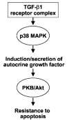
Fig. 9. Schematic representation of pro-survival signaling in mesenchymal cells by TGF-β1.
In the current study, we have demonstrated that TGF-β1 induces rapid activation of p38 MAPK followed by a more delayed activation of the PKB/Akt pathway in lung mesenchymal cells/fibroblasts. Activation of the PI3K/Akt pathway is mediated by p38 MAPK-dependent production of a putative growth factor(s) that functions in an autocrine/paracrine manner. Activation of the PI3K/Akt pathway is sufficient to confer a pro-survival/apoptosis-resistant phenotype to human mesenchymal cells/fibroblasts.

Similar articles

Cited by

References

    1. Massague J. Nat Rev Mol Cell Biol. 2000;1:169–178. - PubMed
    1. Roberts, A. B., and Derynck, R. (2001) Science’s STKE http:/www.stke.org/cgi/content/full/OC_sigtrans;2001/PE43 - PubMed
    1. Border WA, Ruoslahti E. J Clin Investig. 1992;90:1–7. - PMC - PubMed
    1. Grande JP. Proc Soc Exp Biol Med. 1997;214:27–40. - PubMed
    1. Wahl SM, Hunt DA, Wakefield LM, McCartney-Francis N, Wahl LM, Roberts AB, Sporn MB. Proc Natl Acad Sci U S A. 1987;84:5788–5792. - PMC - PubMed

Publication types

MeSH terms