. 2003 Nov 11;100(23):13207-12.
doi: 10.1073/pnas.2135385100. Epub 2003 Oct 23.

The proteome of Saccharomyces cerevisiae mitochondria

Affiliations

The proteome of Saccharomyces cerevisiae mitochondria

Albert Sickmann et al. Proc Natl Acad Sci U S A. .

Abstract

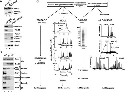
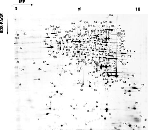
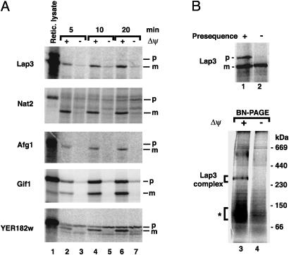
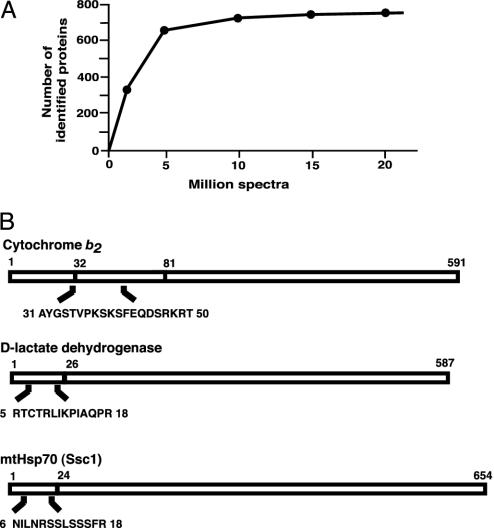
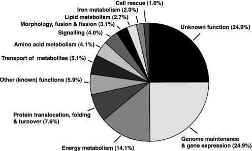
We performed a comprehensive approach to determine the proteome of Saccharomyces cerevisiae mitochondria. The proteins of highly pure yeast mitochondria were separated by several independent methods and analyzed by tandem MS. From >20 million MS spectra, 750 different proteins were identified, indicating an involvement of mitochondria in numerous cellular processes. All known components of the oxidative phosphorylation machinery, the tricarboxylic acid cycle, and the stable mitochondria-encoded proteins were found. Based on the mitochondrial proteins described in the literature so far, we calculate that the identified proteins represent approximately 90% of all mitochondrial proteins. The function of a quarter of the identified proteins is unknown. The mitochondrial proteome will provide an important database for the analysis of new mitochondrial and mitochondria-associated functions and the characterization of mitochondrial diseases.


Figures

Fig. 1.
Fig. 1.
Purification of yeast mitochondria, separation of proteins, and analysis by MS. (A) Subcellular localization of authentic and tagged Tom22. Mitochondria (Mito; 25 μg of protein) and enriched microsomal fractions (25 μg of protein; P100) from WT and Tom22His-10 S. cerevisiae strains were separated by SDS/PAGE and transferred onto poly(vinylidene difluoride) membranes. Immunodecoration was performed with antisera against the indicated proteins and the ECL detection system (Amersham Pharmacia Biotech). Sec61 and Sss1 are subunits of the ER translocon. (B) Highly purified yeast mitochondria. Equal-volume aliquots from total yeast, P12, and highly purified mitochondria were separated by SDS/PAGE and blotted onto poly(vinylidene difluoride) membranes. mALP, mature vacuolar alkaline phosphatase; sALP, soluble vacuolar alkaline phosphatase; Nsp1, subunit of nuclear pore complex; Pex13, peroxisomal biogenesis protein (peroxin) 13; PGK, phosphoglycerate kinase 1. (C) Strategies for protein separation and MS analysis. Four separation methods were used to cover the mitochondrial proteins: isoelectric focusing (IEF) followed by SDS/PAGE (2D PAGE); digestion with four different proteases, followed by multidimensional liquid chromatography (MDLC) and electrospray ionization-MS (ESI-MS); 1D SDS/PAGE, followed by n-LC-MS/MS; generation of a mitochondria-associated fraction by treatment of mitochondria with trypsin or salt, followed by SDS/PAGE and n-LC-MS/MS. Typical examples of separation and spectra are shown.
Fig. 2.
Fig. 2.
Separation of mitochondrial proteins by 2D PAGE. Isolated yeast WT mitochondria were subjected to isoelectric focusing (IEF), followed by SDS/PAGE. The identified protein spots are numbered and listed in Table 2.
Fig. 3.
Fig. 3.
Import of proteins into isolated yeast mitochondria. (A) 35S-labeled precursor proteins were incubated with isolated yeast WT mitochondria in the presence or absence of a membrane potential (ΔΨ) for the indicated times. The samples were treated with proteinase K and separated by SDS/PAGE. Imported proteins were detected by digital autoradiography. Lane 1, nonimported labeled precursor protein in reticulocyte lysate (5% of material added to mitochondria). p, precursor form; m, mature form. (B) Lap3 forms a 300-kDa complex in mitochondria. 35S-labeled Lap3 with or without a presequence (lanes 1 and 2). The precursor form of Lap3 was incubated with isolated mitochondria in the presence or absence of a ΔΨ (lanes 3 and 4). The mitochondria were solubilized by digitonin after the import reaction and separated by blue native PAGE (BN/PAGE). *, Putative dimer of Lap3.
Fig. 4.
Fig. 4.
Identification of mitochondrial proteins and presequences. (A) Number of identified different proteins per million MS spectra. (B) Identification of presequence segments in proteins of the mitochondria-associated fraction. The sequences of the identified peptides found closest to the N terminus are shown. Numbers refer to amino acid positions, starting with the first residue of the presequences. The first and last residues of the mature proteins are indicated. Cytochrome b2 is processed in two steps, after residues 31 and 80 (49).
Fig. 5.
Fig. 5.
Classification of the identified proteins according to function. Protein functions were assigned according to the databases ypd (36), sgd (33), and mitop (43).

References

    1. Schatz, G. (1995) Biochim. Biophys. Acta 1271, 123-126. - PubMed
    1. Scheffler, I. E. (1999) Mitochondria (Wiley, New York).
    1. Lill, R. & Kispal, G. (2000) Trends Biochem. Sci. 25, 352-356. - PubMed
    1. Newmeyer, D. D. & Ferguson-Miller, S. (2003) Cell 112, 481-490. - PubMed
    1. Wallace, D. C. (1999) Science 283, 1482-1488. - PubMed

Publication types