

Repair of oxidative DNA damage by amino acids
- PMID: 14576314
- PMCID: PMC275458
- DOI: 10.1093/nar/gkg816
Repair of oxidative DNA damage by amino acids
Abstract
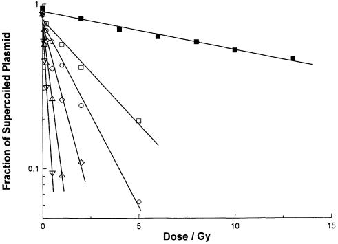
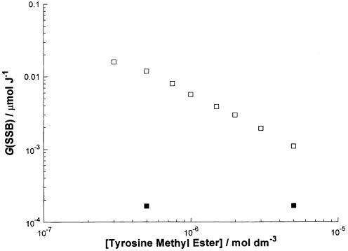
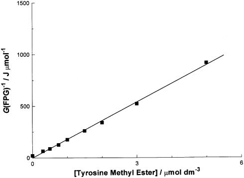
Guanyl radicals, the product of the removal of a single electron from guanine, are produced in DNA by the direct effect of ionizing radiation. We have produced guanyl radicals in DNA by using the single electron oxidizing agent (SCN)2-, itself derived from the indirect effect of ionizing radiation via thiocyanate scavenging of OH. We have examined the reactivity of guanyl radicals in plasmid DNA with the six most easily oxidized amino acids cysteine, cystine, histidine, methionine, tryptophan and tyrosine and also simple ester and amide derivatives of them. Cystine and histidine derivatives are unreactive. Cysteine, methionine, tyrosine and particularly tryptophan derivatives react to repair guanyl radicals in plasmid DNA with rate constants in the region of approximately 10(5), 10(5), 10(6) and 10(7) dm3 mol(-1) s(-1), respectively. The implication is that amino acid residues in DNA binding proteins such as histones might be able to repair by an electron transfer reaction the DNA damage produced by the direct effect of ionizing radiation or by other oxidative insults.
Figures
Similar articles
-
Peptide repair of oxidative DNA damage.Biochemistry. 2004 May 4;43(17):5102-8. doi: 10.1021/bi030232l. Biochemistry. 2004. PMID: 15109269
-
Reaction of guanyl radicals in plasmid DNA with biological reductants: chemical repair of DNA damage produced by the direct effect of ionizing radiation.Int J Radiat Biol. 2001 Nov;77(11):1095-108. doi: 10.1080/09553000110069119. Int J Radiat Biol. 2001. PMID: 11683980
-
Involvement of proton transfer in the reductive repair of DNA guanyl radicals by aniline derivatives.Org Biomol Chem. 2005 Mar 7;3(5):917-23. doi: 10.1039/b418681h. Epub 2005 Feb 7. Org Biomol Chem. 2005. PMID: 15731879
-
Peroxynitrite reactivity with amino acids and proteins.Amino Acids. 2003 Dec;25(3-4):295-311. doi: 10.1007/s00726-003-0018-8. Epub 2003 Sep 26. Amino Acids. 2003. PMID: 14661092 Review.
-
The Two Faces of the Guanyl Radical: Molecular Context and Behavior.Molecules. 2021 Jun 9;26(12):3511. doi: 10.3390/molecules26123511. Molecules. 2021. PMID: 34207639 Free PMC article. Review.
Cited by
-
Microbial cells can cooperate to resist high-level chronic ionizing radiation.PLoS One. 2017 Dec 20;12(12):e0189261. doi: 10.1371/journal.pone.0189261. eCollection 2017. PLoS One. 2017. PMID: 29261697 Free PMC article.
-
Damage clusters after gamma irradiation of a nanoparticulate plasmid DNA peptide condensate.Radiat Environ Biophys. 2012 Mar;51(1):43-52. doi: 10.1007/s00411-011-0388-3. Epub 2011 Oct 2. Radiat Environ Biophys. 2012. PMID: 21964719 Free PMC article.
-
Targeted and Off-Target (Bystander and Abscopal) Effects of Radiation Therapy: Redox Mechanisms and Risk/Benefit Analysis.Antioxid Redox Signal. 2018 Nov 20;29(15):1447-1487. doi: 10.1089/ars.2017.7267. Epub 2018 Mar 22. Antioxid Redox Signal. 2018. PMID: 29350049 Free PMC article. Review.
-
The Role of Amino Acids in Non-Enzymatic Antioxidant Mechanisms in Cancer: A Review.Metabolites. 2023 Dec 31;14(1):28. doi: 10.3390/metabo14010028. Metabolites. 2023. PMID: 38248831 Free PMC article. Review.
-
Chemical and Colloidal Dynamics of MnO2 Nanosheets in Biological Media Relevant for Nanosafety Assessment.Small. 2020 May;16(21):e2000303. doi: 10.1002/smll.202000303. Epub 2020 Mar 19. Small. 2020. PMID: 32191401 Free PMC article.
References
-
- Becker D. and Sevilla,M.D. (1993) The chemical consequences of radiation damage to DNA. Adv. Radiat. Biol., 17, 121–180.
-
- O'Neill P. and Fielden,E.M. (1993) Primary free radical processes in DNA. Adv. Radiat. Biol., 17, 53–120.
-
- Hildenbrand K. and Schulte-Frohlinde,D. (1990) ESR spectra of radicals of single and double stranded DNA in aqueous solution. Implications for OH induced strand breakage. Free Radic. Res. Commun., 11, 195–206. - PubMed
-
- Wolf P., Jones,G.D.D., Candeias,L.P. and O’Neill,P. (1993) Induction of strand breaks in polyribonucleotides and DNA by the sulphate radical anion. Role of electron loss centers as precursors of strand breakage. Int. J. Radiat. Biol., 64, 7–18. - PubMed
