. 2003 Oct 24;302(5645):618-22.
doi: 10.1126/science.1089904.

Experimental models of primitive cellular compartments: encapsulation, growth, and division

Affiliations

Experimental models of primitive cellular compartments: encapsulation, growth, and division

Martin M Hanczyc et al. Science. .

Abstract

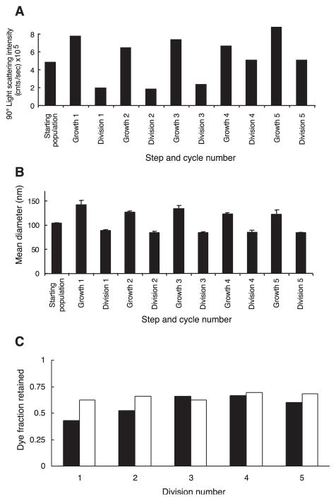
The clay montmorillonite is known to catalyze the polymerization of RNA from activated ribonucleotides. Here we report that montmorillonite accelerates the spontaneous conversion of fatty acid micelles into vesicles. Clay particles often become encapsulated in these vesicles, thus providing a pathway for the prebiotic encapsulation of catalytically active surfaces within membrane vesicles. In addition, RNA adsorbed to clay can be encapsulated within vesicles. Once formed, such vesicles can grow by incorporating fatty acid supplied as micelles and can divide without dilution of their contents by extrusion through small pores. These processes mediate vesicle replication through cycles of growth and division. The formation, growth, and division of the earliest cells may have occurred in response to similar interactions with mineral particles and inputs of material and energy.


Figures

Fig. 1
Fig. 1
Turbidity curves showing vesicle formation over time. Fatty acid micelles were added at 2 min to a buffered solution (0.2 M Na+-bicine, pH 8.5) with or without suspended particles as indicated. (A) Myristoleate (10 mM) added to buffer with 0.6 mg/mL montmorillonite (triangles) or to buffer only (diamonds). (B) Effect of varying concentrations of montmorillonite on vesicle formation. Buffer only (diamonds), 0.02 mg/mL (squares), 0.1 mg/mL (circles), 0.5 mg/mL (triangles). (C) Vesicle formation from different fatty acids in the presence of montmorillonite (0.5 mg/mL, filled shapes) or in buffer alone (open shapes). 10 mM palmitoleate (circles), 10 mM oleate (triangles). (D) Effect of different surfaces on vesicle formation from 10 mM myristoleate micelles. 0.02 mg/mL montmorillonite (squares), 0.02 mg/mL gray Zeeospheres (triangles), 0.02 mg/mL white Zeeospheres (circles), buffer alone (diamonds). (E) Initial rate of vesicle formation [from (B)]versus montmorillonite concentration.
Fig. 2
Fig. 2
Light micrographs of particles and vesicles. Particles visualized with Nomarski optics (A, C, E, and G) and dye-labeled vesicles visualized by fluorescence (B, D, F, H, I, and J). (A and B) Gray Zeeospheres encapsulated within myristoleate vesicles. Arrowheads indicate Zeeospheres in (A) and vesicles containing the Zeeospheres in (B). (C and D) Close-up image of gray Zeeospheres encapsulated within a vesicle. (E and F) Montmorillonite associated with myristoleate vesicles. (G and H) Montmorillonite encapsulated within myristoleate vesicles (arrowhead indicates vesicle containing clay particles). Size bar for (A) to (H), 5 microns. (I) Montmorillonite coated with Cy3-labeled RNA (red) trapped inside dye-labeled vesicles (green). (J) Cy3-labeled RNA (red) in solution encapsulated within labeled vesicle (green). Size bar for (I) and (J), 1 micron.
Fig. 3
Fig. 3
Growth of myristoleate vesicles in response to feeding with one equivalent of myristoleate micelles over a 4-hour period. Changes in vesicle size were followed by (A) dynamic light scattering (DLS) (three replicates) and (B) physical separation by flow-field fractionation coupled with size analysis by multiangle laser-light scattering (FFF-MALLS). RMS, root mean square.
Fig. 4
Fig. 4
FRET assay for membrane growth. (A) Standard curve of normalized Fdon/Facc versus ln- (fatty acid/dye) in myristoleate vesicles (three replicates). (B) Analysis of ln(fatty acid/dye) for the initial vesicle population (before growth), a control population of doubly labeled vesicles 4 hours after the addition of one volume of equimolar unlabeled vesicles (three replicates), grown vesicle population (three replicates), and for comparison the expected ratio assuming 100% fatty acid incorporation into the preexisting vesicle population during growth.
Fig. 5
Fig. 5
Vesicle division by extrusion. Panels show size-exclusion chromatography column profiles of 40 mM myristoleate vesicles with 20 mM encapsulated calcein grown to double their surface area at a constant concentration for 4 hours [(A) 100% of dye encapsulated in vesicles] and subsequently divided 24 hours after the beginning of growth [(B) 43% of dye encapsulated in vesicles].
Fig. 6
Fig. 6
Multiple cycles of growth and division. (A) 90° light-scattering intensities in counts per second, (B) DLS mean diameters (mean and standard deviation of four measurements), and (C) observed (solid bars) and expected (open bars) dye loss for divided populations during each of five cycles (expected values based on DLS diameters before and after division).

Comment in

References

    1. Gebicki JM, Hicks M. Nature. 1973;243:232. - PubMed
    1. Gebicki JM, Hicks M. Chem Phys Lipids. 1976;16:142. - PubMed
    1. Hargreaves WR, Deamer DW. Biochemisty. 1978;17:3759. - PubMed
    1. Apel CL, Deamer DW, Mautner MN. Biochim Biophys Acta. 2002;1559:1. - PubMed
    1. Monnard PA, Apel CL, Kanavarioti A, Deamer DW. Astrobiology. 2002;2:139. - PubMed

Publication types

MeSH terms