

Discoid meniscus in children: treatment and outcome
- PMID: 14577707
- PMCID: PMC3211703
Discoid meniscus in children: treatment and outcome
Abstract
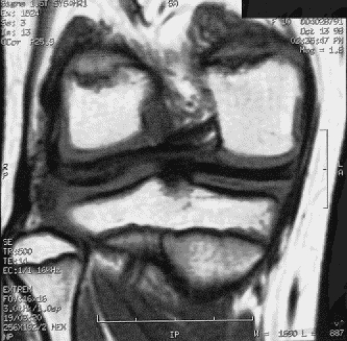
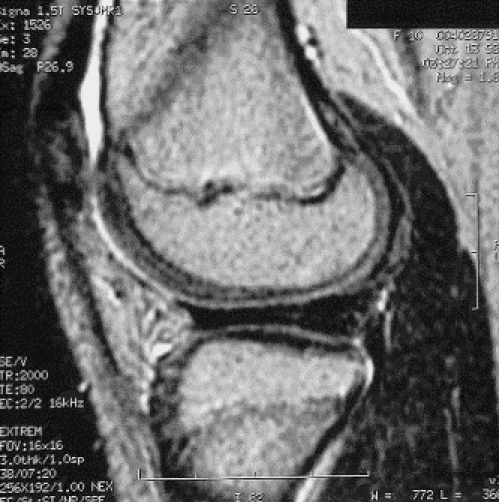
Introduction: Discoid meniscus is an atavistic anomaly in which the meniscus of the knee, predominantly the lateral meniscus, is discoid rather than semilunar in shape. The abnormality is diagnosed relatively infrequently and may even go unrecognized or be untreated. Treatment has consisted of either partial or complete meniscectomy performed either arthroscopically or by open arthrotomy. Our purpose was to examine the outcome of treatment for discoid meniscus in children.
Methods: Since 1974, 34 children, including 2 with bilateral involvement, have been treated for discoid meniscus at the Children's Hospital of Eastern Ontario, a major pediatric referral centre. The average age of the children at the time of surgery was 11 years 4 months (range from 6 yr 2 mo-18 yr 6 mo). The average follow-up was 3 years (range from 1 mo-22 yr). The lateral meniscus was affected in 35 of the 36 knees. Treatment consisted of partial resection in 19 cases, complete resection in 13; 4 did not undergo resection.
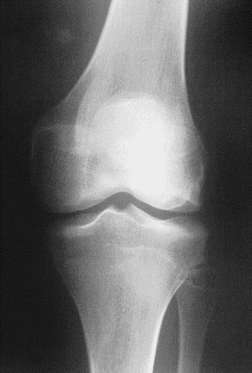
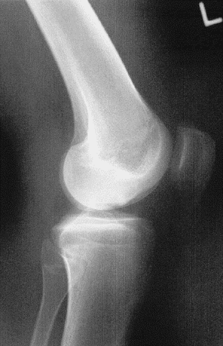
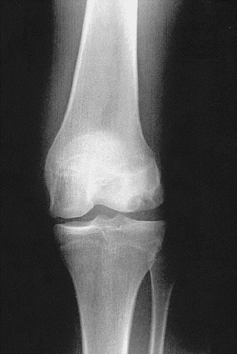
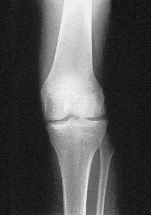
Results: There were 16 excellent, 10 good, 6 fair and 4 poor results at the time of most recent follow-up. In 2 cases degenerative changes were evident radiologically.
Conclusion: Partial resection of discoid menisci is preferable in children, but in complete dislocation of the entire menisci, total removal is necessary.
Introduction: Le ménisque discoïde est une anomalie atavique en raison de laquelle le ménisque du genou, et surtout le ménisque latéral, prend une forme discoïde plutôt que semi-lunaire. Ce diagnostic est relativement rare et il arrive même que l'anomalie ne soit pas identifiée ou traitée. Le traitement a consisté en une méniscectomie partielle ou complète réalisée par arthroscopie ou par athrotomie ouverte. Nous voulions analyser le résultat du traitement du ménisque discoïde chez les enfants.
Méthodes: Depuis 1974, 34 enfants, dont 2 étaient atteints des deux côtés, ont été traités pour un ménisque discoïde au Centre hospitalier pour enfants de l'est de l'Ontario, grand centre de référence pédiatrique. Au moment de l'intervention chirurgicale, les enfants avaient en moyenne 11 ans et 4 mois (intervalle de 6 ans et 2 mois à 18 ans et 6 mois). Le suivi moyen s'est établi à 3 ans (intervalle d'un mois à 22 ans). Le ménisque latéral était atteint dans 35 des 36 genoux. Le traitement a consisté en une résection partielle dans 19 cas, une résection complète dans 13 et 4 sujets n'ont pas subi de résection.
Résultats: Il y avait 16 résultats excellents, 10 bons, 6 moyens et 4 médiocres au moment du suivi le plus récent. Dans deux cas, l'examen radiologique a révélé la présence de changements dégénératifs évidents.
Conclusion: La résection partielle des ménisques discoïdes est préférable chez les enfants, mais dans un cas de luxation complète de tout le ménisque, la résection totale s'impose.
Figures

References
-
- Young RB. The external semilunar cartilage as a complete disc. In: Cleland J, MacKay JY, Young RB, editors. Memoirs and memoranda in anatomy. Vol. 1. London: Williams and Norgate; 1889. p. 179.
-
- Cave EF, Staples OS. Congenital discoid meniscus of cause of internal derangement of the knee. Am J Surg 1941;54:371-6.
-
- Barthel T, Pesch R, Lippert MJ, Lutz G. Arthroskopische Behandlung des lateralen Scheibenmeniskus.Arthroskopie 1995;8:12-8.
-
- Ikeuchi H. Arthroscopic treatment of the discoid lateral meniscus: technique and long-term results. Clin Orthop 1982;167:19-28. - PubMed
Publication types
MeSH terms
LinkOut - more resources
Full Text Sources