Function and structure of pressurized and perfused porcine carotid arteries: effects of in vitro balloon angioplasty
- PMID: 14578174
- PMCID: PMC1892418
- DOI: 10.1016/S0002-9440(10)63533-X
Function and structure of pressurized and perfused porcine carotid arteries: effects of in vitro balloon angioplasty
Abstract
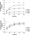
In this report we describe the application of an in vitro pressure-perfusion system for study of functional/structural changes after in vitro balloon dilation injury. Pig carotid arteries were perfused at P = 100 mm Hg and Q = 100 ml/min, balloon angioplastied (BA), and cultured under these hemodynamic conditions for 4 or 8 days (n = 5 BA and 6 controls for each time point). To assess endothelial function, outer diameter changes in response to bradykinin (BK) were measured daily. Remodeling was determined from the shift in pressure-passive diameter relation, as obtained after papaverine addition. Arterial samples were processed for histology. Control arteries showed spontaneous tone, BK-induced relaxation, and inward remodeling that was more pronounced at day 8 (ratio end-to-start passive diameter at P = 100 mm Hg, 0.69 +/- 0.04; P < 0.001) than at day 4 (0.85 +/- 0.03, P = 0.03). Intimal hyperplasia was detectable in these control vessels at day 8 with accumulation of alpha-smooth muscle actin-positive cells around the lumen. Angioplasty caused ruptures and dissections and abolished tone that returned after 5 days of perfusion along with BK-dependent relaxation. No significant inward remodeling or intimal hyperplasia was observed at day 8 after angioplasty. Thus, BA inhibits remodeling, which occurs after in vitro perfusion of conductance arteries.
Figures


References
-
- : American Heart Association: 2002 Heart and Stroke Statistical Update. 2001. American Heart Association Dallas
-
- Narins CR, Holmes DRJ, Topol EJ: A call for provisional stenting: the balloon is back. Circulation 1998, 97:1298-1305 - PubMed
-
- Lafont A, Faxon D: Why do animal models of post-angioplasty restenosis sometimes poorly predict the outcome of clinical trials? Cardiovasc Res 1998, 39:50-59 - PubMed
-
- Teirstein PS: Fulfilling the promise of percutaneous angioplasty. Circulation 2000, 102:2674-2676 - PubMed
-
- Kakuta T, Usui M, Coats WDJ, Currier JW, Numano F, Faxon DP: Arterial remodeling at the reference site after angioplasty in the atherosclerotic rabbit model. Arterioscler Thromb Vasc Biol 1998, 18:47-51 - PubMed
Publication types
MeSH terms
LinkOut - more resources
Full Text Sources

