Recessive epidermolysis bullosa simplex phenotype reproduced in vitro: ablation of keratin 14 is partially compensated by keratin 17
- PMID: 14578178
- PMCID: PMC1892449
- DOI: 10.1016/S0002-9440(10)63537-7
Recessive epidermolysis bullosa simplex phenotype reproduced in vitro: ablation of keratin 14 is partially compensated by keratin 17
Abstract
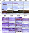
Recessive epidermolysis bullosa simplex (REBS) is characterized by generalized cutaneous blistering in response to mechanical trauma. This results from fragility of the basal keratinocytes that lack keratin tonofilaments because of homozygote null mutation in the keratin 14 gene. REBS patients display in addition focal dyskeratotic skin lesions with histology of epidermolytic hyperkeratosis (EHK) and tonofilament clumping in the suprabasal layers of the epidermis. In this study we examined whether it is possible to mimic in vitro the bullous and dyskeratotic cellular phenotype. For this purpose, fibroblasts from nondyskeratotic (K14-/-) and dyskeratotic (K14-/-) skin of a REBS patient and fibroblasts from a healthy donor (K14+/+) were isolated and incorporated into collagen matrices. Subsequently, fresh biopsies originating from the nondyskeratotic and dyskeratotic skin of the patient and from a healthy donor were placed onto the collagen matrices and cultured at the air-liquid interface. Epidermal morphogenesis was evaluated on the basis of tissue morphology and the expression of a series of keratins. The results of the present study indicate that basal cell vacuolization in REBS can be mimicked in vitro but not the EHK. Fibroblasts seem to play an important regulatory role in establishing the REBS phenotype. These findings suggest that wild-type fibroblasts may enhance the stability of K14-/- keratinocytes in vitro.
Figures


Similar articles
-
In vitro characteristics of early epidermal progenitors isolated from keratin 14 (K14)-deficient mice: insights into the role of keratin 17 in mouse keratinocytes.J Cell Physiol. 1999 Sep;180(3):409-21. doi: 10.1002/(SICI)1097-4652(199909)180:3<409::AID-JCP12>3.0.CO;2-V. J Cell Physiol. 1999. PMID: 10430181
-
A human keratin 10 knockout causes recessive epidermolytic hyperkeratosis.Hum Mol Genet. 2006 Apr 1;15(7):1133-41. doi: 10.1093/hmg/ddl028. Epub 2006 Feb 27. Hum Mol Genet. 2006. PMID: 16505000
-
A missense mutation in the rod domain of keratin 14 associated with recessive epidermolysis bullosa simplex.Nat Genet. 1993 Apr;3(4):327-32. doi: 10.1038/ng0493-327. Nat Genet. 1993. PMID: 7526933
-
Mutations in the genes for epidermal keratins in epidermolysis bullosa and epidermolytic hyperkeratosis.Arch Dermatol. 1993 Dec;129(12):1571-7. Arch Dermatol. 1993. PMID: 7504434 Review.
-
Clinical heterogeneity in recessive epidermolysis bullosa due to mutations in the keratin 14 gene, KRT14.Clin Exp Dermatol. 2008 Nov;33(6):689-97. doi: 10.1111/j.1365-2230.2008.02858.x. Epub 2008 Aug 16. Clin Exp Dermatol. 2008. PMID: 18713255 Review.
Cited by
-
Epidermal growth factor receptor activation and inhibition in 3D in vitro models of normal skin and human cutaneous squamous cell carcinoma.Cancer Sci. 2012 Dec;103(12):2120-6. doi: 10.1111/cas.12026. Epub 2012 Oct 26. Cancer Sci. 2012. PMID: 22974223 Free PMC article.
-
Revertant mosaicism in genodermatoses.Cell Mol Life Sci. 2017 Jun;74(12):2229-2238. doi: 10.1007/s00018-017-2468-2. Epub 2017 Feb 6. Cell Mol Life Sci. 2017. PMID: 28168442 Free PMC article. Review.
-
Skin Disease Models In Vitro and Inflammatory Mechanisms: Predictability for Drug Development.Handb Exp Pharmacol. 2021;265:187-218. doi: 10.1007/164_2020_428. Handb Exp Pharmacol. 2021. PMID: 33387068
-
Epidermolysis bullosa simplex: a paradigm for disorders of tissue fragility.J Clin Invest. 2009 Jul;119(7):1784-93. doi: 10.1172/JCI38177. Epub 2009 Jul 1. J Clin Invest. 2009. PMID: 19587453 Free PMC article. Review.
-
Disruption of epidermal specific gene expression and delayed skin development in AP-2 gamma mutant mice.Dev Biol. 2008 May 1;317(1):187-95. doi: 10.1016/j.ydbio.2008.02.017. Epub 2008 Feb 21. Dev Biol. 2008. PMID: 18353300 Free PMC article.
References
-
- Bonifas JM, Rothman AL, Epstein EH, Jr: Epidermolysis bullosa simplex: evidence in two families for keratin gene abnormalities. Science 1991, 254:1202-1205 - PubMed
-
- Lane EB, Rugg EL, Navsaria H, Leigh IM, Heagerty AH, Ishida-Yamamoto A, Eady RA: A mutation in the conserved helix termination peptide of keratin 5 in hereditary skin blistering. Nature 1992, 356:244-246 - PubMed
-
- Chan Y, Anton-Lamprecht I, Yu Q, Jäckel A, Zabel B, Ernst JP, Fuchs E: A human keratin 14 “knockout”: the absence of K14 leads to severe epidermolysis bullosa simplex and a function for an intermediate filament protein. Genes Dev 1994, 8:2574-2587 - PubMed
Publication types
MeSH terms
Substances
LinkOut - more resources
Full Text Sources
Research Materials
Miscellaneous

