. 2003 Nov;163(5):1801-15.
doi: 10.1016/S0002-9440(10)63540-7.

Abnormalities of basement membrane on blood vessels and endothelial sprouts in tumors

Affiliations

Abnormalities of basement membrane on blood vessels and endothelial sprouts in tumors

Peter Baluk et al. Am J Pathol. 2003 Nov.

Abstract

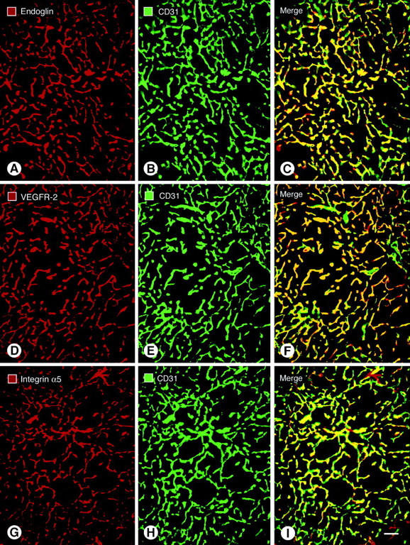
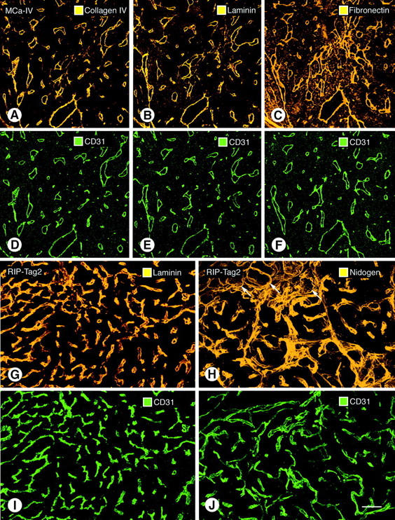
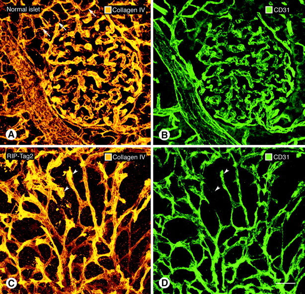
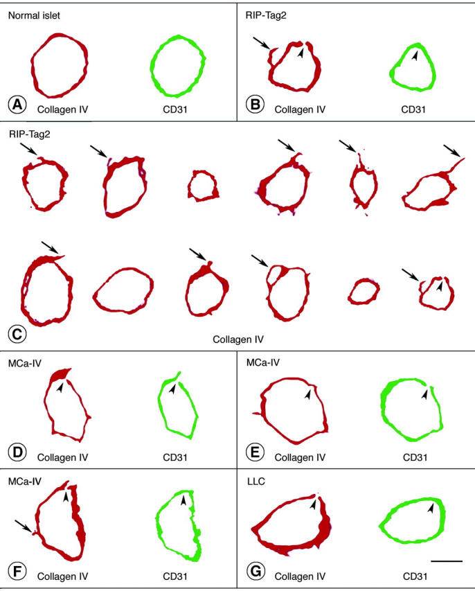
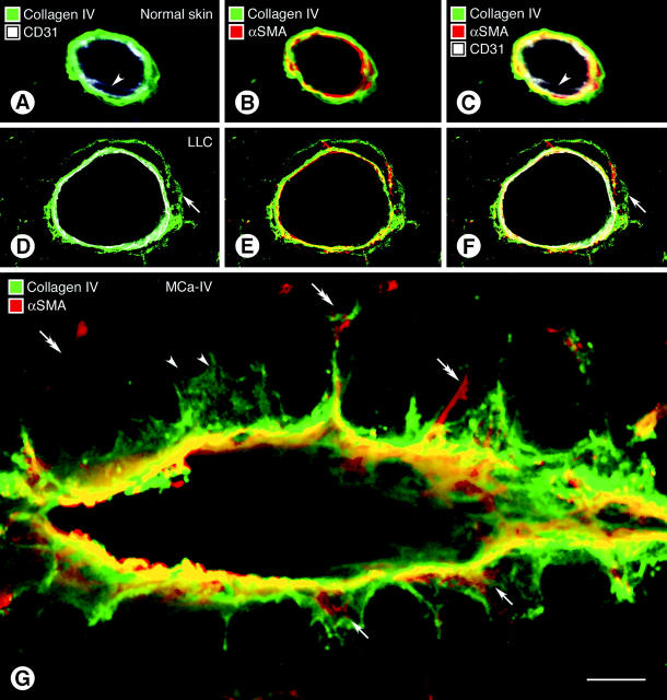
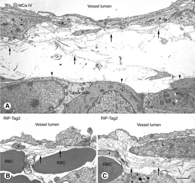
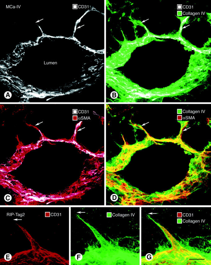
Often described as incomplete or absent, the basement membrane of blood vessels in tumors has attracted renewed attention as a source of angiogenic and anti-angiogenic molecules, site of growth factor binding, participant in angiogenesis, and potential target in cancer therapy. This study evaluated the composition, extent, and structural integrity of the basement membrane on blood vessels in three mouse tumor models: spontaneous RIP-Tag2 pancreatic islet tumors, MCa-IV mammary carcinomas, and Lewis lung carcinomas. Tumor vessels were identified by immunohistochemical staining for the endothelial cell markers CD31, endoglin (CD105), vascular endothelial growth factor receptor-2, and integrin alpha5 (CD49e). Confocal microscopic studies revealed that basement membrane identified by type IV collagen immunoreactivity covered >99.9% of the surface of blood vessels in the three tumors, just as in normal pancreatic islets. Laminin, entactin/nidogen, and fibronectin immunoreactivities were similarly ubiquitous on tumor vessels. Holes in the basement membrane, found by analyzing 1- micro m confocal optical sections, were <2.5 micro m in diameter and involved only 0.03% of the vessel surface. Despite the extensive vessel coverage, the basement membrane had conspicuous structural abnormalities, including a loose association with endothelial cells and pericytes, broad extensions away from the vessel wall, and multiple layers visible by electron microscopy. Type IV collagen-immunoreactive sleeves were also present on endothelial sprouts, supporting the idea that basement membrane is present where sprouts grow and regress. These findings indicate that basement membrane covers most tumor vessels but has profound structural abnormalities, consistent with the dynamic nature of endothelial cells and pericytes in tumors.


Figures

Figure 1.
Figure 1.
Fluorescence micrographs showing blood vessels in RIP-Tag2 tumors double-stained for the endothelial cell markers endoglin (A); VEGFR-2 (D); or integrin alpha5 (G), shown in red; and CD31 immunoreactivity (B, E, H), shown in green. CD31 immunoreactivity primarily co-localizes with the other markers, but the overlap is not perfect (C, F, I). The most complete co-localization, indicated by yellow fluorescence in the merged image, is found with endoglin (C) and VEGFR-2 (F). Some vessels marked by CD31 have little or no integrin alpha5 immunoreactivity, as indicated by green fluorescence in the merged image (I). Few vessels lack CD31 immunoreactivity, as indicated by red fluorescence in merged images (C, F, I). Scale bar, 50 μm (A–I).
Figure 2.
Figure 2.
Confocal microscopic images of vascular basement membrane stained by type IV collagen (A), laminin (B), and fibronectin (C) immunoreactivities in sequential sections of MCa-IV tumor. All three basement membrane proteins co-localize with CD31 immunoreactivity in the same sections (D–F). Type IV collagen immunoreactivity is primarily restricted to blood vessels in these tumors, but laminin and fibronectin immunoreactivities are also present elsewhere in the tumors. Laminin (G) and entactin/nidogen (H) immunoreactivities and corresponding CD31 immunoreactivity (I, J) are shown associated with blood vessels in RIP-Tag2 tumors. Arrows in H mark exocrine pancreatic acini outlined by entactin/nidogen immunoreactivity. Scale bars: 170 μm (A–F); 50 μm (G–J).
Figure 3.
Figure 3.
Confocal microscopic images of type IV collagen immunoreactivity of basement membrane (A, C) and CD31 immunoreactivity of endothelial cells (B, D) in normal pancreatic islet of wild-type mouse (A, B) and in RIP-Tag2 tumor (C, D). In both cases, all CD31-immunoreactive vessels co-localize with type IV collagen immunoreactivity. Tumor vessels are more irregular and tortuous but still have a uniform envelopment of type IV collagen immunoreactivity. Arrowheads mark tumor vessels where type IV collagen staining (C) is broader or more prominent than the corresponding CD31 immunoreactivity (D), suggestive of a loose association with endothelial cells. Arrows in A point to exocrine pancreatic acini outlined by type IV collagen immunoreactivity. Scale bar, 50 μm (A–D).
Figure 4.
Figure 4.
Type IV collagen (red) and CD31 immunoreactivities (rat clone MEC13.3 anti-mouse, green) viewed in two-dimensional projections of 1-μm confocal microscopic optical cross-sections of vessels in normal pancreatic islets (A), RIP-Tag2 tumors (B, C), MCa-IV breast carcinomas (D–F), and Lewis lung carcinoma (G). Physical section thickness, 80 μm. Type IV collagen immunoreactivity completely envelops most vessels. Arrows mark regions of type IV collagen immunoreactivity that extend beyond the CD31-immunoreactive perimeter of tumor vessels. Arrowheads mark tiny defects (0.3 to 2.5 μm in diameter) in type IV collagen immunoreactivity and/or CD31 immunoreactivity of vessels in the three types of tumor, as detailed in Table 1 ▶ . There are several reasons why CD31 immunoreactivity of endothelial cells appears as a single band instead of two discrete (luminal and abluminal) plasma membranes in these images. To visualize the immunofluorescence in 1-μm optical sections of endothelial cells, the confocal signal was amplified to a point plasma membranes appear wider than they really are. The apparent widening of the membranes is exaggerated by slight tilting from vertical of vessels not cut precisely in cross-section. The separation of the two membranes is less than the thickness of the optical sections (except in the region of the nucleus) and is of the same order of magnitude as the resolution of the ×40 objective lens. Because of the interaction of these factors, the width of the signal from both membranes blurs the space between them, and they fuse into one fluorescent band. Scale bars: 5 μm (A, B, G); 7.5 μm (C); 45 μm (D–F).
Figure 5.
Figure 5.
Confocal microscopic images comparing wall structure of 5-μm-thick optical sections of vessels in normal skin (A–C) and Lewis lung carcinoma (D–F, LLC) triple-stained for CD31, α-SMA, and type IV collagen immunoreactivities. Type IV collagen staining in the normal venule is closely apposed to CD31-immunoreactive endothelial cells (A) and α-SMA-immunoreactive pericytes (B). All three markers form a tight sandwich (C). Arrowheads mark endothelial cell nucleus. In the tumor vessel a layer of type IV collagen immunoreactivity extends well beyond the endothelial cells (D–F, arrows) and in some regions co-localizes with α-SMA-positive pericytes (E). A composite view (F) shows that the cell layers and basement membrane are separated by spaces (black). CD31 staining in some images (A, C, D, F) appears somewhat diffuse and not precisely limited to the plasma membrane for the reasons mentioned in the legend for Figure 4 ▶ . In G, multiple basement membrane abnormalities are shown in an oblique section through a vessel in MCa-IV breast carcinoma, double-stained for type IV collagen (green) and α-SMA (red) immunoreactivities, with regions of greatest overlap appearing yellow. Type IV collagen immunoreactivity spreads loosely from the vessel wall and penetrates the surrounding stroma (black). Extensions of type IV collagen immunoreactivity envelop α-SMA-immunoreactive pericytes in some regions (arrows) but not in others (arrowheads). Some α-SMA-immunoreactive cell processes extend away from the vessel wall (double arrows). Scale bar, 10 μm (A–G).
Figure 6.
Figure 6.
Transmission electron micrographs of blood vessels in MCa-IV mammary carcinoma (A) and RIP-Tag2 tumor (B, C) with basement membrane (arrows) that is structurally abnormal but provides complete vessel coverage. Endothelial cells line the vessel lumen. Pericytes are marked by asterisks. Basement membrane is loosely associated with endothelial cells and pericytes and in some regions extends away from the vessel wall (A, arrows). Some regions of the vascular basement membrane are multilayered (B and C, arrows). By contrast, the basement membrane on tumor cell clusters is uniformly thin, compact, and closely apposed to tumor cells (A, arrowheads). RBC, extravasated erythrocytes. Scale bar, 2.5 μm (A); 1 μm (B, C).
Figure 7.
Figure 7.
Relationship of basement membrane (type IV collagen immunoreactivity, green), endothelial cells (CD31 immunoreactivity, white), and pericytes (α-SMA immunoreactivity, red) in vascular sprouts (arrows) in 80-μm-thick sections of MCa-IV tumor (A–D) and RIP-Tag2 tumor (E–G). Vessel in A–D cut obliquely. The tip of CD31-stained sprouts (A, E) ends short of the distal-most extension of type IV collagen immunoreactivity (B, F, G) and α-SMA immunoreactivity (C). However, type IV collagen staining coincides with the α-SMA immunoreactivity of pericyte processes on the sprouts (D). Scale bar, 50 μm (A–D); 5 μm (E–G).

References

    1. Pasqualini R, Arap W, McDonald DM: Probing the structural and molecular diversity of tumor vasculature. Trends Mol Med 2002, 8:563-571 - PubMed
    1. St. Croix B, Rago C, Velculescu V, Traverso G, Romans KE, Montgomery E, Lal A, Riggins GJ, Lengauer C, Vogelstein B, Kinzler KW: Genes expressed in human tumor endothelium. Science 2000, 289:1197-1202 - PubMed
    1. Folkman J: Angiogenesis-dependent diseases. Semin Oncol 2001, 28:536-542 - PubMed
    1. Jain RK: Normalizing tumor vasculature with anti-angiogenic therapy: a new paradigm for combination therapy. Nat Med 2001, 7:987-989 - PubMed
    1. Warren BA: The vascular morphology of tumors. Peterson HE eds. Tumor Blood Circulation. 1979:pp 1-48 CRC Press Inc., Boca Raton

Publication types

MeSH terms