Comparative Study
. 2003 Nov;163(5):1859-71.
doi: 10.1016/S0002-9440(10)63545-6.

In vivo interrogation of the molecular display of atherosclerotic lesion surfaces

Affiliations
Comparative Study

In vivo interrogation of the molecular display of atherosclerotic lesion surfaces

Cheng Liu et al. Am J Pathol. 2003 Nov.

Abstract

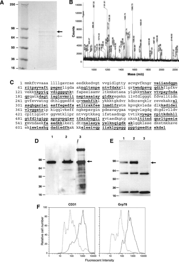
The endothelial surface of atherosclerotic lesions of ApoE knockout mice was interrogated by in vivo biopanning with a phage-displayed constrained peptidyl library. Through repeated biopanning, 103 peptidyl sequences were identified, many are homologous to known proteins. The sequence CAPGPSKSC contains motifs that are shared by 9.7% of selected peptides. On phage or as a synthetic peptide, this constrained peptide selectively bound to atherosclerotic lesion surfaces of ApoE knockout mice in vivo and of human atherosclerotic lesions ex vivo. A cell-surface protein of approximately 82 kd recognized by this peptide was affinity-purified and determined by mass spectrometry analysis as glucose-regulated protein 78 (Grp78), indicating the surprising presence of this endoplasmic reticulum chaperone on the endothelial cell surface of atherosclerotic lesions. Peptides that mimicked binding functions of their homologues were demonstrated with three peptides homologous to tissue inhibitor of metalloproteinase-2 (TIMP-2), ie, CNHRYMQMC, CNQRHQMSC, and CNNRSDGMC. Phage carrying CNHRYMQMC bound to atherosclerotic lesion endothelium of ApoE knockout mice in vivo. The three peptides bound to endothelial cells in a dose-dependent manner and were inhibited by TIMP-2 protein. These peptides provide a set of probes to interrogate the cell surface repertoire associated with atherogenesis and thrombotic complications.


Figures

Figure 1.
Figure 1.
Aortas from atherosclerotic ApoE mice injected with 1011 phage either expressing CAPGPDSKSC or not (control phage). Binding of phage to aorta was visualized with biotinylated anti-phage antibody and streptavidin-linked peroxidase activation of DAB. A: The aorta of an atherosclerotic ApoE knockout mouse injected with CAPGPSKSC phage. Phage binding to the lesions is readily visualized (red-brown). B: The aorta from an atherosclerotic ApoE knockout mouse in which lesions are clearly evident. This mouse was infused with control phage and there is no evident association of phage with the lesions. C: The normal aorta from a BALB/c mouse infused with the CAPGPSKSC phage. No association of phage with the aortic surface is observed suggesting the CAPGPSKSC target molecule is absent or not present in detectable quantity. D: The aorta of a young ApoE knockout mouse on standard chow diet and without atherosclerotic lesions when infused with CAPGPSKSC phage, no localization was detectable indicating that the CAPGPSKSC target molecule is not expressed simply as a result of ApoE gene inactivation.
Figure 2.
Figure 2.
A: An aorta from an atherosclerotic ApoE knockout mouse and infused with the synthetic biotinylated CAPGPSKSC peptide. Peptide binding to the lesions is visualized (red-brown) and replicates the phage binding. B: A histological section of a typical atherosclerotic lesion of the aorta of an atherosclerotic ApoE knockout mouse. Endothelium overlying the atherosclerotic lesion was identified with anti-CD31 antibody and labeled dark brown (arrows). C: An adjacent section. The binding of this peptide is most intense to the endothelium overlying the atherosclerotic lesion (yellow arrows), but negative on endothelial cells not directly overlying lesions (white arrows). D: A positive control with all endothelial cells recognized by an anti-CD31 antibody. E: A negative control and autofluorescence resulted from elastic tissue in the vessel wall. Original magnifications, ×200.
Figure 3.
Figure 3.
Image of an atherosclerotic lesion of a human iliac artery segment. The tissue before staining is shown at the left. Binding of biotinylated CAPGPSKSC peptide to the lesion is demonstrated after application to the lumenal surface of the vessel (right). The binding of phage to the lesion was visualized with enzyme-linked streptavidin conversion of DAB substrate to the brownish red product. The association of CAPGPSKSC peptide to the lesion, but not to nonatherosclerotic surface of this and other vessels (not shown), indicates that a human homologue of the target molecule is present. Lesions are outlined.
Figure 4.
Figure 4.
Identification of a target molecule in mouse endothelial cells (bEND.3). The total protein lysate (lane 1) or membrane preparation (lane 2) was separated by SDS-polyacrylamide gel electrophoresis then transferred to nitrocellulose membrane. The latter was probed with biotinylated peptide CAPGPSKSC. Two sharp bands are observed on a Western blot of a whole cell lysate of mouse endothelial cell line (bEND.3). The protein bands bound by the CAPGPSKSC peptide were ∼82 kd and ∼120 kd. The sharpness of the detected bands suggests that these target proteins are not glycosylated. The ∼82-kd protein was also more abundant in cellular fractions enriched for membrane proteins by Triton X-114 extraction, consistent with a membrane localization of this protein.
Figure 5.
Figure 5.
Characterization of Grp78 as a target for CAPGPSKSC peptide. A: The Coomassie-stained gel after protein affinity purification on an immobilized CAPGPSKSC column. The black arrow indicates a protein band of ∼82 kd and the gray arrow indicates a proteolytic product of the ∼82-kd protein. B: The cryptic peptide mixture of the ∼82-kd protein band analyzed by matrix-assisted laser desorption/ionization mass spectrometer. C: The matched cryptic peptide sequences of the ∼82-kd protein band in mouse Grp78 (shown in bold and underlined). D: The Western blotting of the enriched bEND.3 membrane extract with antibodies against 1) the N-terminus of Grp78, 2) the C-terminus of Grp78, and 3) by peptide-ligand blot using biotinylated CAPGPSKSC. E: Immunoprecipitation of the enriched bEND.3 membrane extract with antibodies against 1) the N-terminus of Grp78, 2) the C-terminus of Grp78, 3) a control antibody (anti-tissue factor). Blotting was performed using biotinylated CAPGPSKSC peptide. The lower bands are immunoglobulin fragments in the reaction. F: The flow cytometry analysis of cell surface Grp78 molecules on bENd.3 cells using anti-Grp78 polyclonal antisera. Note a shift of fluorescent intensity in cells stained with anti-Grp78 antisera, similar to cells stained with anti-CD31 antibody.
Figure 6.
Figure 6.
Images of phage bound to aortic valves. These aortic valves were from ApoE knockout mice with atherosclerotic lesions. The mice were infused with 1011 pfu of phage carrying the CNQRHQMSC sequence. In A, a H&E-stained section of aortic valve from an atherosclerotic ApoE knockout mouse is shown. Note the aortic valve is thickened by the presence of atherosclerotic lesions. In B, the endothelial cells report red with biotinylated rat anti-mouse CD 31 antibody and the associated phage report green with anti-phage antibody. Co-localization reports yellow. In experiments performed with control phage, no phage association was evident as shown in C. These data indicate that the CNQRHQMSC phage can bind to atherosclerotic lesion-involved endothelial surfaces. Original magnifications, ×100.
Figure 7.
Figure 7.
The three synthetic peptides with homology to TIMP-2 bind to endothelial cells in a dose-dependent manner (top). Binding of peptides (0.5 μmol/L) was inhibited by purified TIMP-2 protein (0.05 μmol/L) (bottom), indicating that the peptides mimicking TIMP-2 binding are consistent with a novel binding site for TIMP-2 protein on the surface of these cells. The scrambled control peptide did not bind.

References

    1. Libby P: Changing concepts of atherogenesis. J Intern Med 2000, 247:349-358 - PubMed
    1. Faggiotto A, Ross R: Studies of hypercholesterolemia in the nonhuman primate. II. Fatty streak conversion to fibrous plaque. Arteriosclerosis 1984, 4:341-356 - PubMed
    1. Rosenfeld ME, Tsukada T, Gown AM, Ross R: Fatty streak initiation in Watanabe heritable hyperlipemic and comparably hypercholesterolemic fat-fed rabbits. Arteriosclerosis 1987, 7:9-23 - PubMed
    1. Ross R: The pathogenesis of atherosclerosis: a perspective for the 1990s. Nature 1993, 362:801-809 - PubMed
    1. Ross R: Atherosclerosis: current understanding of mechanisms and future strategies in therapy. Transplant Proc 1993, 25:2041-2043 - PubMed

Publication types

MeSH terms