Comparative Study
. 2003 Nov;163(5):1949-60.
doi: 10.1016/S0002-9440(10)63553-5.

Non-small-cell lung cancer molecular signatures recapitulate lung developmental pathways

Affiliations
Comparative Study

Non-small-cell lung cancer molecular signatures recapitulate lung developmental pathways

Alain C Borczuk et al. Am J Pathol. 2003 Nov.

Abstract

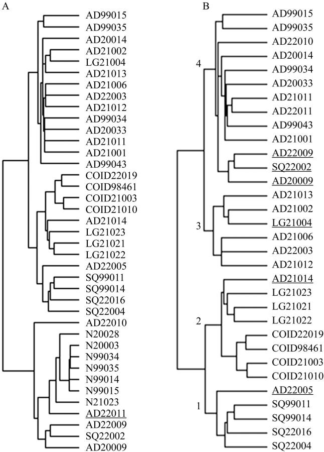
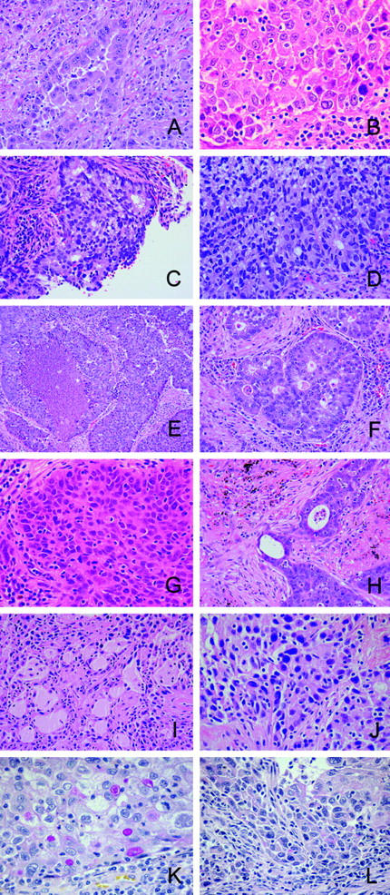
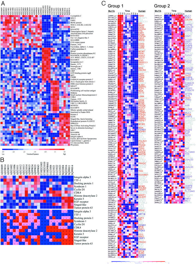
Current paradigms hold that lung carcinomas arise from pleuripotent stem cells capable of differentiation into one or several histological types. These paradigms suggest lung tumor cell ontogeny is determined by consequences of gene expression that recapitulate events important in embryonic lung development. Using oligonucleotide microarrays, we acquired gene profiles from 32 microdissected non-small-cell lung tumors. We determined the 100 top-ranked marker genes for adenocarcinoma, squamous cell, large cell, and carcinoid using nearest neighbor analysis. Results were validated by immunostaining for 11 selected proteins using a tissue microarray representing 80 tumors. Gene expression data of lung development were accessed from a publicly available dataset generated with the murine Mu11k genome microarray. Self-organized mapping identified two temporally distinct clusters of murine orthologues. Supervised clustering of lung development data showed large-cell carcinoma gene orthologues were in a cluster expressed in pseudoglandular and canalicular stages whereas adenocarcinoma homologues were predominantly in a cluster expressed later in the terminal sac and alveolar stages of murine lung development. Representative large-cell genes (E2F3, MYBL2, HDAC2, CDK4, PCNA) are expressed in the nucleus and are associated with cell cycle and proliferation. In contrast, adenocarcinoma genes are associated with lung-specific transcription pathways (SFTPB, TTF-1), cell adhesion, and signal transduction. In sum, non-small-cell lung tumors histology gene profiles suggest mechanisms relevant to ontogeny and clinical course. Adenocarcinoma genes are associated with differentiation and glandular formation whereas large-cell genes are associated with proliferation and differentiation arrest. The identification of developmentally regulated pathways active in tumorigenesis provides insights into lung carcinogenesis and suggests early steps may differ according to the eventual tumor morphology.


Figures

Figure 1.
Figure 1.
Unsupervised clustering hierarchical dendrograms are determined by histology and morphology. A: Dendrogram of entire set of 39 specimens. Underlined specimen AD22011 is a well-differentiated bronchioloalveolar carcinoma that segregates within clade of nonmalignant specimens. B: Dendrogram of 32 tumor specimens. Restricting the clustering to tumor specimens, lung carcinomas cluster into four clades that are associated with histology. Placement of outlier specimens (underlined) with histology classification dissimilar from other members in the clade can be explained by examination of histological sections.
Figure 2.
Figure 2.
Photomicrographs of underlined tumors from dendrogram in Figure 1B ▶ . A and B: AD22005 from clade 1 segregated with squamous carcinomas. A: A gland-forming adenocarcinoma containing small areas of squamous differentiation (B), which accounted for less than 10% of the tumor’s morphology. C and D: AD21014 from clade 2 segregated with large-cell carcinomas. C: Gland-forming areas that comprised more than 10% of the tumor’s morphology. The remainder of the tumor showed poor differentiation (D) similar to the large-cell tumors. E and F: LG21004 from clade 3 segregated with adenocarcinomas. The tumor morphology showed large-cell features of organoid growth with central necrosis (E) and strong reactivity for neuroendocrine markers synaptophysin and chromogranin (data not shown). A minority of the tumor showed cribiform gland structures (F). G–L: Clade 4 contained one squamous cell carcinoma SQ22002 (G) with rare fields showing glandular differentiation (H) and two adenocarcinomas AD22009 and AD20009, which showed a majority of adenocarcinoma pattern (I and K, respectively) as well as areas of squamous differentiation (J and L, respectively). A–J, H&E stain; K and L, mucicarmine stain. Original magnifications: ×50 (E); ×100 (A, C, D, F, H, I); ×150 (B, G, J–L).
Figure 3.
Figure 3.
Histological class gene marker sets determined by nearest neighbor supervised clustering. A: Nearest neighbor analysis with a t-test metric was used to identify and rank 100 gene markers for each non-small-cell lung cancer histology class relative to other classes (Table 2) ▶ . The top 15 markers per histology are shown. For each gene, red indicates a high level of expression relative to the median and blue indicates a low level of expression relative to the median. B: Validation of gene marker sets with immunohistochemistry. Eleven proteins for which antibodies previously used on paraffin sections were commercially available were selected. Top: Supervised clustering diagram excerpts from Figure 3A ▶ . Bottom: Antibody reactivity for each sample. For each protein, blue, pink, and red indicate a score of 0 (negative), 1 (low positive), and 2 (high positive), respectively. C: Nearest neighbor analysis was used to classify and rank Mu11k gene markers associated with two temporally associated gene groups. Genes were filtered to remove 22 murine genes without a human orthologue in the histology marker sets. Shown are expression profiles for group 1 (left) and for group 2 (right) genes that were more highly associated with group distinction than 95% of random permutations of the group labels (P < 0.05). Lung development stages represented by group 1 (days E12 to E18)- and group 2 (postnatal D1 to adult)-expressed genes are pseudoglandular/canalicular and terminal sac/alveolar, respectively. Each row shows gene expression throughout time for murine genes. The corresponding human histology gene orthologues for large-cell carcinoma are indicated in red, adenocarcinoma in blue. Other gene labels for carcinoid and squamous carcinomas are indicated in gray.

References

    1. Jemal A, Murray T, Samuels A, Ghafoor A, Ward E, Thun MJ: Cancer statistics, 2003. CA Cancer J Clin 2003, 53:5-26 - PubMed
    1. Ferlay J, Bray F, Pisani P, Parkin DM: Globocan 2000: Cancer Incidence, Mortality and Prevalence. 2001. IARC Press Lyon
    1. Travis WD, Colby TV, Corrin B, Shimosato Y, Brambilla E: Histological typing of lung and pleural tumors. World Health Organization International Histological Classification of Tumours. 1999. Springer-Verlag New York
    1. Stellman SD, Muscat JE, Thompson S, Hoffmann D, Wynder EL: Risk of squamous cell carcinoma and adenocarcinoma of the lung in relation to lifetime filter cigarette smoking. Cancer 1997, 80:382-388 - PubMed
    1. Reya T, Morrison SJ, Clarke MF, Weissman IL: Stem cells, cancer, and cancer stem cells. Nature 2001, 414:105-111 - PubMed

Publication types

MeSH terms