. 2003 Nov 11;100(23):13680-5.
doi: 10.1073/pnas.1835674100. Epub 2003 Oct 24.

Axonal transport of eukaryotic translation elongation factor 1alpha mRNA couples transcription in the nucleus to long-term facilitation at the synapse

Affiliations

Axonal transport of eukaryotic translation elongation factor 1alpha mRNA couples transcription in the nucleus to long-term facilitation at the synapse

Maurizio Giustetto et al. Proc Natl Acad Sci U S A. .

Abstract

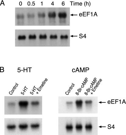
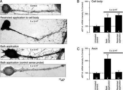
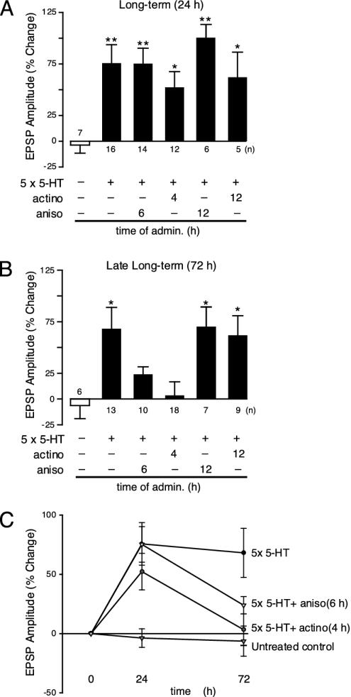
Long-term synaptic plasticity requires both gene expression in the nucleus and local protein synthesis at synapses. The effector proteins that link molecular events in the cell body with local maintenance of synaptic strength are not known. We now show that treatment with serotonin (5-HT) that produces long-term facilitation induces the Aplysia eukaryotic translation elongation factor 1alpha (Ap-eEF1A) as a late gene that might serve this coupling function in sensory neurons. Although the translation factor is induced, it is not transported into axon processes when the stimulation with 5-HT was restricted to the cell body. In contrast, its mRNA is transported when 5-HT was applied to both cell body and synapses. Intracellular injection of antisense oligonucleotides or antibodies that block the induction and expression of Ap-eEF1A do not affect the initial expression of long-term facilitation but do block its maintenance beyond 24 h. The transport of eEF1A protein and its mRNA to nerve terminals suggests that the translation factor plays a role in the local protein synthesis that is essential for maintaining newly formed synapses.


Figures

Fig. 1.
Fig. 1.
Induction of eEF1A mRNA. (A) Time course. Total RNA was extracted from the nervous tissue from untreated Aplysia (controls) or animals treated with 5-HT and probed with 32P-labeled Ap-eEF1A cDNA. As control, the RNA samples were also probed with an S4-ribosomal cDNA that is not affected by 5-HT (21, 44). Radioactivity was quantified densitometrically, and the extent of stimulation was calculated by normalizing eEF1A values with those of S4. (B) (Left) Inhibition of induction by emetine. Total RNA was isolated 6 h after the start of the treatment with 5-HT alone or with 5-HT and emetine, and probed with 32P-labeled Ap-eEF1A cDNA (Upper) or S4 cDNA (Lower). The 7-fold (± 0.4; n = 6; P < 0.001) induction of eEF1A mRNA observed with 5-HT (Student's paired t test) was inhibited by emetine. (Right) Induction by 8-Br-cAMP. Total RNA from ganglia treated with 8-Br-cAMP and probed with 32P-labeled Ap-eEF1A cDNA (Upper) or S4 cDNA (Lower). This 5.5 (± 0.5-fold; P < 0.01; n = 6) induction was also blocked by emetine.
Fig. 2.
Fig. 2.
5-HT stimulation is necessary for transport of eEF1A mRNA into neurites. Images of cultured neurons making synaptic contact with L7 motor neurons are shown. Sensory neurons were stimulated with five pulses of 5-HT, and the amount of Ap-EF1A mRNA was measured as pixel intensity by in situ hybridization using a digoxigenin-labeled antisense mRNA probe (see Supporting Methods). (A) Neurites of the untreated (control) cell showed no staining. Local application of 5-HT to the sensory neuron's cell body resulted in the staining of the cell body and proximal axon but not of the distal portion of the axon. A strong signal for eEF1A mRNA, detected by the antisense probe, is evident in the cell body and axon of the neuron stimulated by bath application of 5-HT. Cells treated with 5-HT were not stained when hybridized with a control sense probe. Quantitation of the mean pixel intensity corresponding to the somatic (B) and axonal (C) eEF1A mRNA staining was done in sensory cells stimulated by bath-applied 5-HT and compared with cells where 5-HT stimulation was restricted to the cell body. The RNA signal in each group was normalized to the mean signal obtained for the untreated group. Bath application of 5-HT increased the amount of eEF1A mRNA in the cell body (B) and in axons (C) (**, P < 0.01, treated versus untreated controls). In contrast, application of 5-HT restricted to the cell body increased the amount of staining only in the cell body (B, **, P < 0.01, treated versus untreated controls; ANOVA and Neuman-Keul's multiple range test), and not in the axon (C).
Fig. 3.
Fig. 3.
Regional distribution of Ap-eEF1A determined by immunoblotting. (A) An antipeptide antibody raised against the predicted amino acid sequence (Ser-440—Lys-459) recognized a single Mr 50,000 component in extracts of Aplysia nervous tissue (see Supporting Methods). Preimmune serum was not reactive. Immunoreactivity was abolished when the antibodies were preincubated with the peptide immunogen. (B) Immunoblotting of the extracts from the sensory neuron's cell body and neuropil show that eEF1A immunoreactivity is present in both compartments (see Supporting Methods). Reprobing the same blot with antisynaptotagmin antibodies (Santa Cruz Biotechnology) revealed the enrichment of synaptic protein in the neuropil fraction compared with that of the cell body. Cell bodies and neuropil were separated under a light microscope from ganglia treated with 2 M NaCl and 50% propylene glycol (1:1, vol/vol) and kept at -20°C for 5 h (45), which renders the solid ganglia translucent while preserving the cytological structure and thereby aids in dissection. (C) Induction of eEF1A protein. Extracts were made from sensory neurons treated with 5-HT or controls and immunoblotted (Upper). The same samples were probed with antiubiquitin antibody (Sigma) (Lower). Because the amount of ubiquitin and ubiquitin conjugates do not change after treatment with 5-HT (46), this serves as control loading control. (D) Quantification of eEF1A immunoreactivity in untreated and 5-HT-treated sensory neurons. The amount of eEF1A is significantly higher (P < 0.01; n = 6, paired t test) in 5-HT-treated sensory neurons.
Fig. 4.
Fig. 4.
A second period of gene expression is required to maintain late-phase synaptic facilitation. EPSPs were recorded from cultured motor neurons. Changes (%) in EPSP amplitude (mean ± SEM) are shown as bar graphs. (A) Anisomycin (aniso, 10 μM applied for 1 h) did not affect the early phase of long-term facilitation (24 h) if administered at 6 or 12 h after treatment with five pulses of 5-HT (**, P < 0.01, ANOVA and Neuman-Keul's multiple range test) nor did actinomycin (actino, 50 μg/ml) administered at 4 or 12 h after treatment with 5-HT at 24 h (*, P < 0.05 and **, P < 0.01). (B) The late phase of long-term facilitation was blocked when anisomycin was applied at 6 h but not at 12 h after the stimulation with 5-HT (*, P < 0.05). Late long-term facilitation was also abolished when actinomycin D was administered 4 h after the five pulses of 5-HT, but was unaffected when transcription was inhibited at 12 h (*, P < 0.05; **, P < 0.01). (C) Effects of blocking transcription and translation on EPSP amplitude. To inhibit translation, actinomycin was added 4 h after the application of five pulses of 5-HT. To block protein synthesis, anisomycin was applied 6 h after the treatment with 5-HT.
Fig. 5.
Fig. 5.
Inhibition of Ap-eEF1A prevents the late but not the early phase of long-term facilitation. EPSPs were recorded from cultured motor neurons. Sensory neurons were injected with antibodies or oligonucleotides or were uninjected. Changes (%) in EPSP amplitude (mean ± SEM) are shown as bar graphs. (A) EPSPs were recorded at 24 h after five 5-min pulses of 5-HT. Anti-eEF1A antibody (Ab) inhibited the early phase if injected 1 h before 5-HT treatment but not at 6 or 12 h after treatment with 5-HT. Injection of preimmune (Pre) serum had no effect. eEF1A antisense oligonucleotide (AS) also had no effect on the induction of long-term facilitation at 24 h if injected 6 h after the 5-HT treatment (**, P < 0.01, ANOVA and Neuman-Keul's multiple range test). (B) EPSPs were recorded at 72 h after five 5-min pulses of 5-HT. Late long-term facilitation was completely blocked when anti-eEF1A antibody was injected 1 h before and 6 h after the 5-HT application. Fully expressed late long-term facilitation was seen with anti-eEF1A antibody injected 12 h after the 5-HT treatment. Injection of preimmune serum had no effect. Injection of eEF1A antisense oligonucleotides at 6 h after the 5-HT treatment also completely blocked late long-term facilitation (*, P < 0.05). (C) EPSPs were recorded at 10 min (short-term facilitation) after one 5-min pulse of 5-HT. Neither the anti-eEF1A antibody nor the antisense oligonucleotide had any effect on short-term facilitation (1 × 5-HT). Preimmune serum and scrambled oligonucleotide (Scr) also had no effect (**, P < 0.01).

Similar articles

Cited by

References

    1. Kandel, E. R. & Schwartz, J. H. (1982) Science 218, 433-443. - PubMed
    1. Kandel, E. R. (2001) Science 294, 1030-1038. - PubMed
    1. Martin, K. C., Barad, M. & Kandel, E. R. (2000) Curr. Opin. Neurobiol. 10, 587-592. - PubMed
    1. Jiang, C. & Schuman, E. M. (2002) Trends Biochem. Sci. 27, 506-513. - PubMed
    1. Kiebler, M. A. & DesGroseillers, L. (2000) Neuron 25, 19-28. - PubMed

Publication types

MeSH terms

LinkOut - more resources