. 2003 Nov;228(3):451-63.
doi: 10.1002/dvdy.10429.

Neurogenic phenotype of mind bomb mutants leads to severe patterning defects in the zebrafish hindbrain

Affiliations

Neurogenic phenotype of mind bomb mutants leads to severe patterning defects in the zebrafish hindbrain

Stephanie Bingham et al. Dev Dyn. 2003 Nov.

Abstract

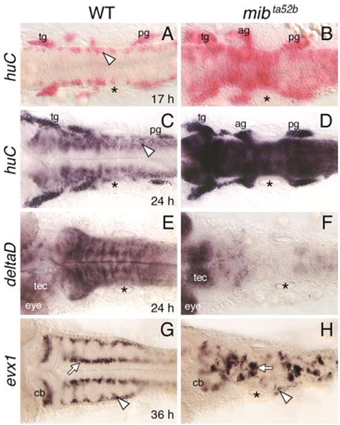
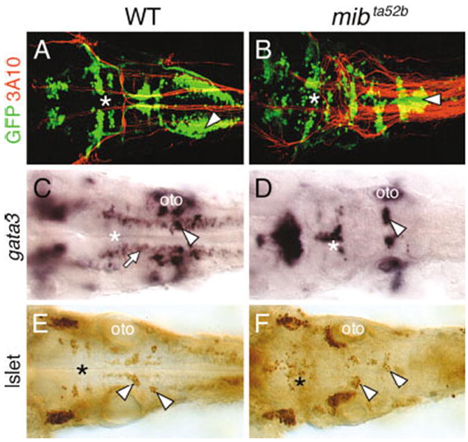
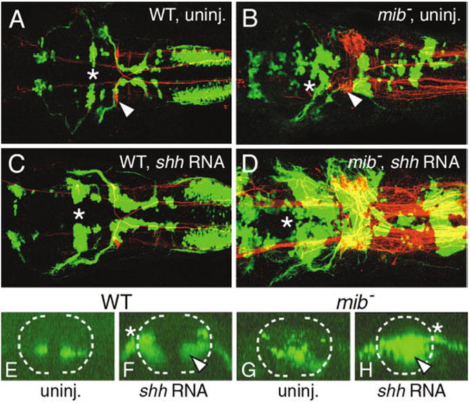
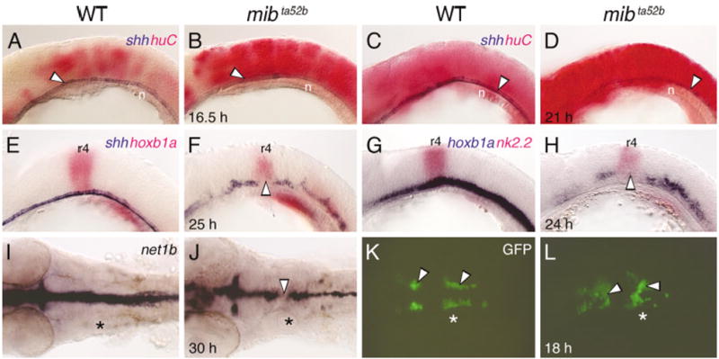
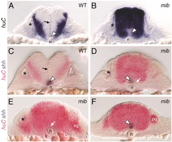
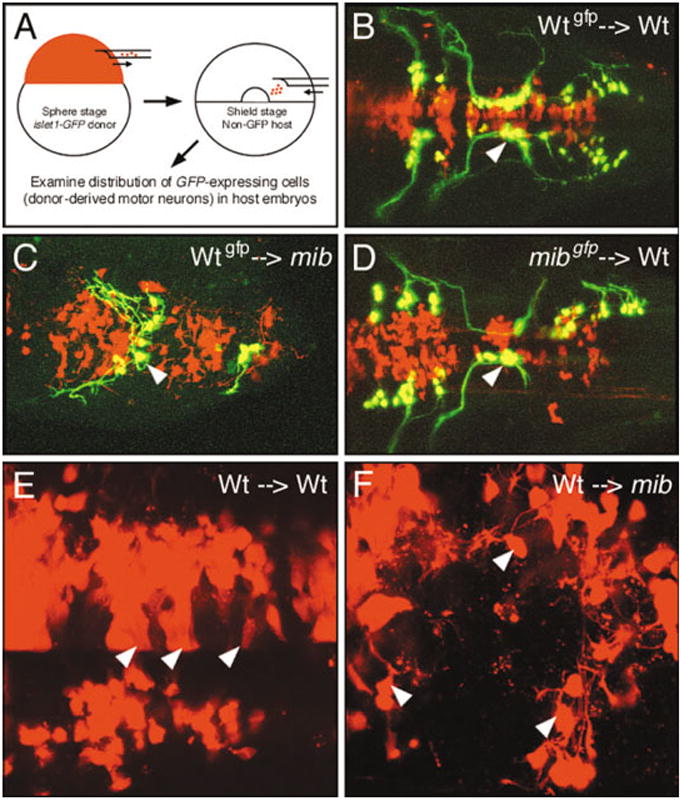
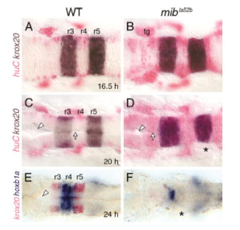
Failure of Notch signaling in zebrafish mind bomb (mib) mutants results in a neurogenic phenotype where an overproduction of early differentiating neurons is accompanied by the loss of later-differentiating cell types. We have characterized in detail the hindbrain phenotype of mib mutants. Hindbrain branchiomotor neurons (BMNs) are reduced in number but not missing in mib mutants. In addition, BMN clusters are frequently fused across the midline in mutants. Mosaic analysis indicates that the BMN patterning and fusion defects in the mib hindbrain arise non-cell autonomously. Ventral midline signaling is defective in the mutant hindbrain, in part due to the differentiation of some midline cells into neural cells. Interestingly, while early hindbrain patterning appears normal in mib mutants, subsequent rhombomere-specific gene expression is completely lost. The defects in ventral midline signaling and rhombomere patterning are accompanied by an apparent loss of neuroepithelial cells in the mutant hindbrain. These observations suggest that, by regulating the differentiation of neuroepithelial cells into neurons, Notch signaling preserves a population of non-neuronal cells that are essential for maintaining patterning mechanisms in the developing neural tube.


Figures

Fig. 1
Fig. 1
Defective neurogenesis in the mind bomb mutant hindbrain. All panels show dorsal views of the hindbrain with anterior to the left. The asterisks in all panels indicate the location of the otocyst. A,B: In a 17 hours postfertilization (hpf) wild-type embryo (WT; A), huC is expressed in small clusters of cells (arrowhead) located at the lateral margins of the hindbrain. In a mib mutant (B), huC-expressing cells are found throughout the hindbrain, with higher densities at some axial levels. C,D: In a 24 hpf wild-type embryo (C), the huC expression domain has expanded medially, and expressing cells are found at all axial levels, including the caudal hindbrain (arrowhead). In a mib mutant (D), the hindbrain at all axial levels is filled with huC-expressing cells. E,F: In a 24 hpf wild-type embryo (E), deltaD is expressed extensively, with higher expression in segmentally reiterated cell clusters. In the mib mutant (F), deltaD expression is greatly reduced or absent in the hindbrain and sharply higher in the tectum (tec) and eye. G,H: In a 36 hpf wild-type embryo (G), evx1 is expressed in commissural neurons (arrowhead), cerebellar neurons (cb), and putative interneurons (arrow). In a mib mutant (H), the evx1-expressing commissural neurons are greatly reduced (arrowhead), while the cerebellar neurons and interneurons (arrow) are disorganized and fused, and reduced in number. ag, acoustic ganglion; cb, cerebellum; tec, tectum; tg, trigeminal ganglion; pg, posterior lateral line ganglion.
Fig. 2
Fig. 2
Branchiomotor neuron (BMN) development is severely disrupted in the mind bomb mutant hindbrain. All panels show dorsal views of the hindbrain with anterior to the left. A and B are composite confocal images and identify GFP-expressing motor neurons in the fluorescein channel and 3A10-labeled Mauthner (M) reticulospinal neurons and axons in the rhodamine channel. The asterisks in all panels indicate the location of the trigeminal motor neurons in rhombomeres 2 and 3. A,B: In a 36 hpf wild-type embryo (WT; A), the BMN clusters are found in their characteristic locations and numbers. The 3A10 antibody-labeled Mauthner cell axons decussate and descend contralaterally into the spinal cord. In a mib mutant (B), The BMNs clusters are disorganized and are variably fused. The supernumerary M cell axons cross the midline and descend contralaterally in a normal manner, whereas the nX motor neurons (arrowhead) in the same region exhibit extensive fusion across the midline. C,D: In a 36 hpf wild-type embryo (C), gata3 is expressed by putative interneurons (arrow) and the nVII (arrowhead) and nV motor neurons (asterisk). In a mib mutant (D), gata3-expressing motor neurons (arrowhead, asterisk) are disorganized and the putative interneurons are absent. E,F: In a 36 hpf mib mutant (F), islet antibody-labeled BMNs (asterisk, arrowheads) are disorganized and variably fused and are reduced in number compared with wild-type siblings (E). oto, otocyst.
Fig. 3
Fig. 3
Shh overexpression induces branchiomotor neurons (BMNs) in the mind bomb mutant hindbrain. A–D show dorsal views of the hindbrain with anterior to the left and are composite confocal images, identifying GFP-expressing motor neurons in the fluorescein channel and 3A10-labeled Mauthner reticulospinal neurons and axons in the rhodamine channel. Asterisks in A–D indicate the location of the trigeminal (nV) motor neurons in rhombomere 2 (r2). E–H show confocal projections (virtual cross-sections) of GFP-expressing cells at the level of r4 and r5, and illustrate the effects of shh overexpression on nVII motor neurons. The broken line marks the outline of the neural tube, with dorsal to the top. The asterisks in F and H indicate the exit point of nVII axons from the neural tube. A: In a control wild-type embryo (WT), one Mauthner (M) neuron (arrowhead) is found on each side in r4 and BMNs are found in characteristic locations and numbers. E: In addition, the nVII motor neuron cell bodies are found in the ventral neural tube. C: In a shh RNA-injected wild-type embryo, the various GFP-expressing BMN clusters contain significantly larger numbers of cells, while M cell number is not affected. F: While most of the ectopic nVII motor neurons are found in the dorsal neural tube, a significant number (arrowhead) is also found in ventral locations. B: In a control mib mutant, there are excess M cells (arrowhead) and the BMNs exhibit the mib mutant phenotype. G: The nVII neurons are distributed randomly in the ventral half of the neural tube. D: Upon shh RNA injection, there is a sharp increase in the various BMN populations, while M cell number is not affected. H: Significantly, many of the ectopic nVII motor neurons (arrowhead) in the mutant are found in the ventral neural tube. The mutant embryo shown in D had an unusually large increase in motor neuron number at all axial levels.
Fig. 4
Fig. 4
Development of ventral neural tube tissue is defective in the mind bomb hindbrain. A–H show lateral views and I–L show dorsal views of the hindbrain, with anterior to the left. Asterisks (I–L) indicate the location of the otocyst. A–D: At 16.5 hours postfertilization (hpf; A,B) and 21 hpf (C,D), shh-expressing floor plate cells (arrowheads) are found in a continuous row in wild-type (WT) and mib mutant embryos, even though huC-expressing cells are greatly increased in the mutant (B, D). E,F: In a 24 hpf mib mutant (F), shh-expressing floor plate cells are disorganized and slightly reduced in number compared with a wild-type sibling (E), with an absence of expressing cells in rhombomere 4 (r4, arrowhead), which is identified by hoxb1a expression. G,H: In a 24 hpf mib mutant (H), expression of nk2.2 in the ventral neural tube is greatly reduced compared with a wild-type embryo (G), with an absence of expression in r4 (arrowhead). I,J: In a 30 hpf mib mutant (J), net1b expression is reduced at all axial levels compared with wild-type (I), with a gap of nonexpression in r4 (arrowhead). K,L: In an 18 hpf wild-type embryo (K), GFP-expressing nV and nVII motor neurons (arrowheads) are found bilaterally in r2 (nV neurons) and in a longitudinal column spanning r4 and r5 (nVII neurons). In a mib mutant (L), the motor neuron clusters (arrowheads) are disorganized, with considerable fusion across the midline. n, notochord.
Fig. 5
Fig. 5
Distribution of neural cells in the mind bomb mutant hindbrain. All panels show cross-sections of the hindbrain in 24 hours postfertilization embryos at the level of rhombomere 5 (A–D), rhombomere 4 (E), and the caudal hindbrain (F). Asterisks in A–E identify the otocyst. A,C: In wild-type (WT) embryos, the huC-expressing cells are found at the lateral margins of the neural tube, outside the ventricular zone along the middle of the neural tube (arrows). The ventral midline contains floor plate cells (arrowheads) that express shh (C). B,D: In mib mutants, the entire neural tube is filled with huC-expressing cells, except at the ventral midline (arrowhead in B), which contains shh-expressing cells (arrowhead in D) and other cells that express neither shh nor huC (arrow in D). The ventricular zone is absent in mutant embryos. E: In this mutant, huC-expressing cells (arrow) occupy the ventral midline immediately dorsal to the notochord, and no shh-expressing cells are found. F: In this mutant, the ventral midline cells coexpress shh and huC (arrowhead). ag, acoustic ganglion; n, notochord; pg, posterior lateral line ganglion.
Fig. 6
Fig. 6
The mind bomb branchiomotor neuron (BMN) phenotype arises non–cell autonomously. B–F show dorsal views of the hindbrain with anterior to the left. In B–F, donor-derived cells are labeled red with rhodamine–dextran. In B–D, donor-derived BMNs are labeled yellow due to colocalization of GFP expression and rhodamine–dextran. A: The transplantation procedure. B: In a control experiment, wild-type (Wt) cells that differentiate into motor neurons (arrowhead) in a wild-type host are correctly organized into characteristic clusters. C: When donor-derived wild-type cells differentiate into motor neurons in a mib mutant host, the wild-type neurons (arrowhead) and their axons are loosely organized and straddle the midline, the characteristic mutant phenotype. D: When donor-derived mib mutant cells differentiate into motor neurons in a wild-type host, the mutant neurons (arrowhead) and their axons exhibit wild-type organization. E: In a wild-type host, many donor-derived cells exhibit a columnar morphology (arrowheads), suggestive of neuroepithelial cells. F: In a mib host, most donor-derived cells are round with short processes (arrowheads), suggestive of neurons.
Fig. 7
Fig. 7
Rhombomere patterning is established but not maintained in mind bomb mutants. All panels show dorsal views with anterior to the left. Asterisks identify the otocyst. A–D: At 16.5 hours postfertilization (hpf; A,B) and 20 hpf (C,D), krox20 is expressed normally in rhombomeres 3 and 5 (r3 and r5) in wild-type (WT) and mutant embryos, even though huC-expressing cells are greatly increased in the mutant hindbrains (B,D). By 20 hpf, the ventricular surface (arrow) and lumen (arrowhead) are well developed in the wild-type embryo (C) but are poorly developed in the mutant (D). E,F: In a mib mutant (F), krox20 expression in r3 and r5 is absent, while hoxb1a expression in r4 is maintained but greatly reduced, compared with a wild-type sibling (E). In addition, the ventricle (arrowhead) seen in the wild-type embryo is missing in the mutant. tg, trigeminal ganglion.

References

    1. Appel B, Korzh V, Glasgow E, Thor S, Edlund T, Dawid IB, Eisen JS. Motoneuron fate specification revealed by patterned LIM homeobox gene expression in embryonic zebrafish. Development. 1995;121:4117–4125. - PubMed
    1. Appel B, Fritz A, Westerfield M, Grunwald DJ, Eisen JS, Riley BB. Delta-mediated specification of midline cell fates in zebrafish embryos. Curr Biol. 1999;9:247–256. - PubMed
    1. Artavanis-Tsakonas S, Rand MD, Lake RJ. Notch signaling: cell fate control and signal integration in development. Science. 1999;284:770–776. - PubMed
    1. Barth KA, Wilson SW. Expression of zebrafish nk22 is influenced by sonic hedgehog/vertebrate hedgehog-1 and demarcates a zone of neuronal differentiation in the embryonic forebrain. Development. 1995;121:1755–1768. - PubMed
    1. Bingham S, Higashijima S, Okamoto H, Chandrasekhar A. The zebrafish trilobite gene is essential for tangential migration of branchiomotor neurons. Dev Biol. 2002;242:149–160. - PMC - PubMed

Publication types

LinkOut - more resources