. 2003 Nov 4;42(43):12654-61.
doi: 10.1021/bi034446e.

Effects of DNA adduct structure and sequence context on strand opening of repair intermediates and incision by UvrABC nuclease

Affiliations

Effects of DNA adduct structure and sequence context on strand opening of repair intermediates and incision by UvrABC nuclease

Yue Zou et al. Biochemistry. .

Abstract

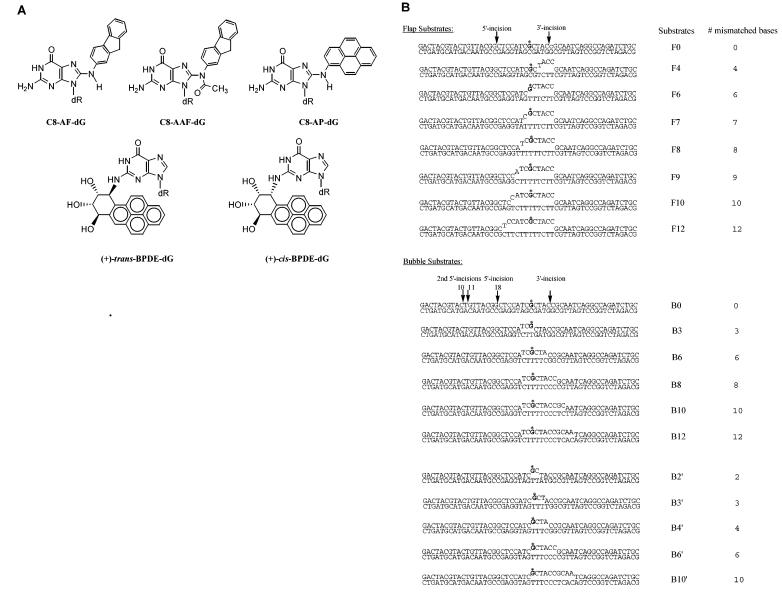
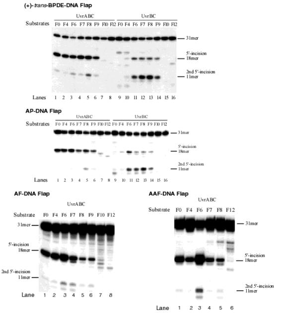
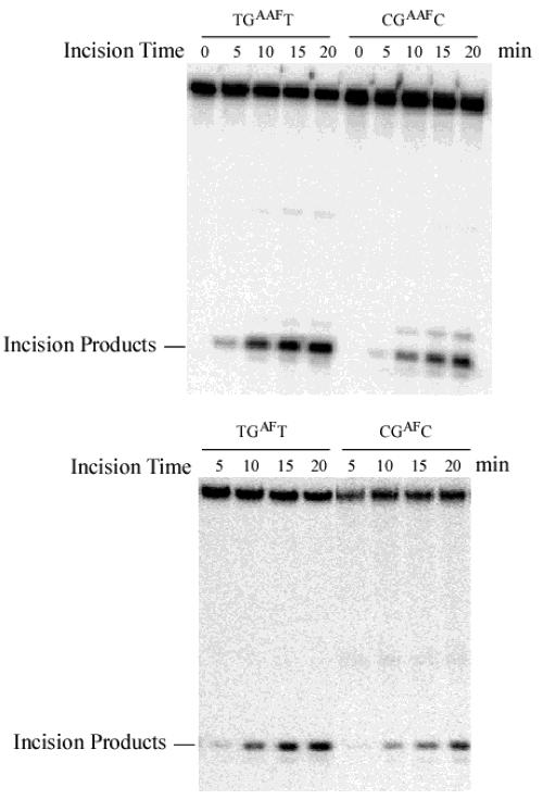
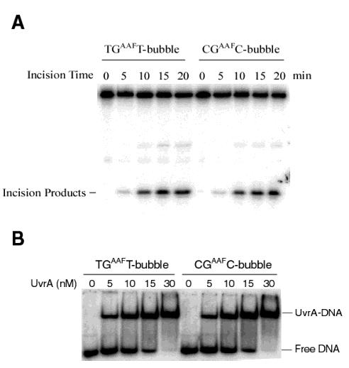
DNA damage recognition of nucleotide excision repair (NER) in Escherichia coli is achieved by at least two steps. In the first step, a helical distortion is recognized, which leads to a strand opening at the lesion site. The second step involves the recognition of the type of chemical modification in the single-stranded region of DNA during the processing of the lesions by UvrABC. In the current work, by comparing the efficiencies of UvrABC incision of several types of different DNA adducts, we show that the size and position of the strand opening are dependent on the type of DNA adducts. Optimal incision efficiency for the C8-guanine adducts of 2-aminofluorene (AF) and N-acetyl-2-aminofluorene (AAF) was observed in a bubble of three mismatched nucleotides, whereas the same for C8-guanine adduct of 1-nitropyrene and N(2)-guanine adducts of benzo[a]pyrene diol epoxide (BPDE) was noted in a bubble of six mismatched nucleotides. This suggests that the size of the aromatic ring system of the adduct might influence the extent and number of bases associated with the opened strand region catalyzed by UvrABC. We also showed that the incision efficiency of the AF or AAF adduct was affected by the neighboring DNA sequence context, which, in turn, was the result of differential binding of UvrA to the substrates. The sequence context effect on both incision and binding disappeared when a bubble structure of three bases was introduced at the adduct site. We therefore propose that these effects relate to the initial step of damage recognition of DNA structural distortion. The structure-function relationships in the recognition of the DNA lesions, based on our results, have been discussed.


Figures

FIGURE 1:
FIGURE 1:
Chemical structure of DNA adducts (A) and the list of substrates (B) used in the present study. The bold G with an asterisk in the sequence (B) represents the adducted base, the arrows indicate the major incision sites, the lowercase base (C) in the top strand of the flap substrate F0 indicates there is a nick between the bases (CG), and the c represents a free 3′ end. Only the flap substrates contain an internal nick or free 3′ end, and the bubble substrates do not. The flap size increases in the direction of 3′ to 5′, while the bubble expands from 5′ to 3′.
FIGURE 2:
FIGURE 2:
Incisions of DNA adducts of flap substrate by UvrABC and UvrBC nucleases. The 5′-terminally labeled flap substrates were incubated with UvrABC (UvrA, 15 nM; UvrB, 250 nM; and UvrC, 100 nM) or UvrBC in the UvrABC buffer at 37 °C for 30 min. The incision products were analyzed on a 12% polyacrylamide sequencing gel. The 31mer represents the intact top strand DNA (labeled), while the 18mer and 10-11mer are the products of normal 5′-incision and second 5′-incisions, respectively. The second 5′-incisions are coupled with and dependent on the normal 5′-incision so that both types of incision products should be combined for determination of incision efficiency. The four different types of adducts, (+)-trans-BPDE-, AP-, AF-, and AAF-DNA were assayed under the same conditions.
FIGURE 3:
FIGURE 3:
Incisions of DNA adducts of bubble substrate by UvrABC nucleases. The 5′-terminally labeled DNA substrates with varying bubble sizes were incubated with UvrABC (UvrA, 15 nM; UvrB, 250 nM; and UvrC, 100 nM) in the UvrABC buffer at 37 °C for 30 min. The incision products were analyzed on a 12% polyacrylamide sequencing gel. The 50mer represents the intact DNA substrate. The 18mer and 11mer stand for the normal 5′- and the second 5′-incision products, respectively. The second 5′-incisions are coupled with and dependent on the normal 5′-incision so that both types of incision products should be combined for determination of incision efficiency. The four different types of adducts, (+)-trans-BPDE-, AP-, AF-, and AAF-DNA were assayed under the same conditions.
FIGURE 4:
FIGURE 4:
Effect of DNA sequence context at adducts on the incision efficiency of UvrABC. The 5′-terminally labeled DNA substrates adducted with AF and AAF in TG*T and CG*C sequences were incubated with UvrABC (UvrA, 15 nM; UvrB, 250 nM; and UvrC, 100 nM) in the UvrABC buffer at 37 °C for the indicated periods. The incision products were analyzed on a 12% polyacrylamide urea denatureing gel. Incision efficiency or rate was determined for each of the substrates as the slope of a linear regression line of the kinetic data.
FIGURE 5:
FIGURE 5:
Binding of UvrA protein to the AAF-DNA substrates in TG*T and CG*C sequences. The 5′-terminally labeled DNA substrates were incubated with varying concentrations of UvrA at 37 °C for 15 min in the UvrABC buffer without ATP. The binding products were analyzed on a 3.5% native polyacrylamide gel. Free DNA represents the unbound DNA substrates, and the UvrA-DNA represents the protein-DNA complex formation.
FIGURE 6:
FIGURE 6:
Incision and binding of AAF-DNA bubble substrates. (A) The AAF-DNA bubble substrates (B3′, Figure 1B) with the adduct sequences TG*T and CG*C were incised by UvrABC nuclease. The 5′-terminally labeled substrates were incubated with UvrABC (UvrA, 15 nM; UvrB, 250 nM; and UvrC, 100 nM) in the UvrABC buffer at 37 °C for various periods. The incision products were analyzed on a 12% polyacrylamide sequencing gel. (B) Binding of UvrA to the same bubble substrates. The 5′-terminally labeled DNA substrates were incubated with varying concentrations of UvrA at 37 °C for 15 min in the UvrABC buffer without ATP. The binding products were analyzed on a 3.5% native polyacrylamide gel.

Similar articles

Cited by

References

    1. Van Houten B. Microbiol. ReV. 1990;54:18–51. - PMC - PubMed
    1. Sancar A. Annu. ReV. Biochem. 1996;65:43–81. - PubMed
    1. Lindahl T, Wood RD. Science. 1999;286:1897–1905. - PubMed
    1. Orren DK, Sancar A. J. Biol. Chem. 1990;265:15796–15803. - PubMed
    1. Shi Q, Thresher R, Sancar A, Griffith J. J. Mol. Biol. 1992;226:425–432. - PubMed

Publication types

MeSH terms