

Cytoplasmic p21WAF1/CIP1 expression is correlated with HER-2/ neu in breast cancer and is an independent predictor of prognosis
- PMID: 14580260
- PMCID: PMC314414
- DOI: 10.1186/bcr654
Cytoplasmic p21WAF1/CIP1 expression is correlated with HER-2/ neu in breast cancer and is an independent predictor of prognosis
Abstract
Background: HER-2 (c-erbB2/Neu) predicts the prognosis of and may influence treatment responses in breast cancer. HER-2 activity induces the cytoplasmic location of p21WAFI/CIPI in cell culture, accompanied by resistance to apoptosis. p21WAFI/CIPI is a cyclin-dependent kinase inhibitor activated by p53 to produce cell cycle arrest in association with nuclear localisation of p21WAFI/CIPI. We previously showed that higher levels of cytoplasmic p21WAFI/CIPI in breast cancers predicted reduced survival at 5 years. The present study examined HER-2 and p21WAFI/CIPI expression in a series of breast cancers with up to 9 years of follow-up, to evaluate whether in vitro findings were related to clinical data and the effect on outcome.
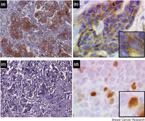
Methods: The CB11 anti-HER2 monoclonal antibody and the DAKO Envision Plus system were used to evaluate HER-2 expression in 73 patients. p21WAFI/CIPI staining was performed as described previously using the mouse monoclonal antibody Ab-1 (Calbiochem, Cambridge, MA, USA).
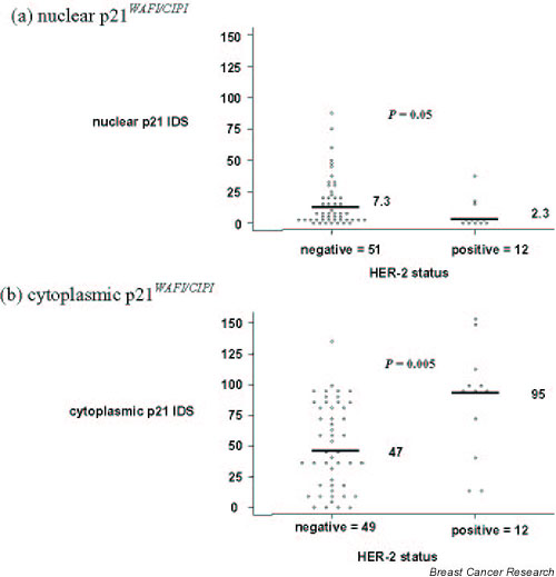
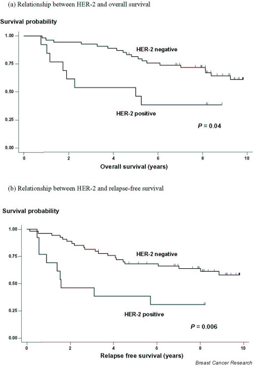
Results: HER-2 was evaluable in 67 patients and was expressed in 19% of cases, predicting reduced overall survival (P = 0.02) and reduced relapse-free survival (P = 0.004; Cox regression model). HER-2-positive tumours showed proportionately higher cytoplasmic p21WAFI/CIPI staining using an intensity distribution score (median, 95) compared with HER-2-negative cancers (median, 47) (P = 0.005). There was a much weaker association between nuclear p21WAFI/CIPI and HER-2 expression (P = 0.05), suggesting an inverse relationship between nuclear p21WAF1/CIP1 and HER-2.
Conclusion: This study highlights a new pathway by which HER-2 may modify cancer behaviour. HER-2 as a predictor of poor prognosis may partly relate to its ability to influence the relocalisation of p21WAFI/CIPI from the nucleus to the cytoplasm, resulting in a loss of p21WAFI/CIPItumour suppressor functions. Cytoplasmic p21WAFI/CIPI may be a surrogate marker of functional HER-2 in vivo.
Figures
Similar articles
-
p14ARF expression in invasive breast cancers and ductal carcinoma in situ--relationships to p53 and Hdm2.Breast Cancer Res. 2004;6(5):R571-85. doi: 10.1186/bcr912. Epub 2004 Jul 29. Breast Cancer Res. 2004. PMID: 15318938 Free PMC article.
-
Phosphorylation/cytoplasmic localization of p21Cip1/WAF1 is associated with HER2/neu overexpression and provides a novel combination predictor for poor prognosis in breast cancer patients.Clin Cancer Res. 2004 Jun 1;10(11):3815-24. doi: 10.1158/1078-0432.CCR-03-0527. Clin Cancer Res. 2004. PMID: 15173090
-
Novel signaling molecules implicated in tumor-associated fatty acid synthase-dependent breast cancer cell proliferation and survival: Role of exogenous dietary fatty acids, p53-p21WAF1/CIP1, ERK1/2 MAPK, p27KIP1, BRCA1, and NF-kappaB.Int J Oncol. 2004 Mar;24(3):591-608. Int J Oncol. 2004. PMID: 14767544
-
Expression patterns of cyclins D1, E and cyclin-dependent kinase inhibitors p21waf1/cip1, p27kip1 in colorectal carcinoma: correlation with other cell cycle regulators (pRb, p53 and Ki-67 and PCNA) and clinicopathological features.Int J Clin Pract. 2008 Nov;62(11):1736-43. doi: 10.1111/j.1742-1241.2006.01105.x. Int J Clin Pract. 2008. PMID: 19143860
-
HER-2/neu (c-erb-B2) gene and protein in breast cancer.Am J Clin Pathol. 1999 Jul;112(1 Suppl 1):S53-67. Am J Clin Pathol. 1999. PMID: 10396301 Review.
Cited by
-
Cooperative interactions of HER-2 and HPV-16 oncoproteins in the malignant transformation of human mammary epithelial cells.Neoplasia. 2005 Aug;7(8):788-98. doi: 10.1593/neo.05106. Neoplasia. 2005. PMID: 16207481 Free PMC article.
-
Verrucarin A alters cell-cycle regulatory proteins and induces apoptosis through reactive oxygen species-dependent p38MAPK activation in the human breast cancer cell line MCF-7.Tumour Biol. 2014 Oct;35(10):10159-67. doi: 10.1007/s13277-014-2286-1. Epub 2014 Jul 16. Tumour Biol. 2014. PMID: 25027398
-
Sustained Supratherapeutic Paclitaxel Delivery Enhances Irreversible Sarcoma Cell Death.Mol Cancer Ther. 2022 Nov 3;21(11):1663-1673. doi: 10.1158/1535-7163.MCT-21-0750. Mol Cancer Ther. 2022. PMID: 36031342 Free PMC article.
-
The novel roles of virus infection-associated gene CDKN1A in chemoresistance and immune infiltration of glioblastoma.Aging (Albany NY). 2021 Feb 17;13(5):6662-6680. doi: 10.18632/aging.202519. Epub 2021 Feb 17. Aging (Albany NY). 2021. PMID: 33621203 Free PMC article.
-
A prognostic index for operable, node-negative breast cancer.Br J Cancer. 2004 May 17;90(10):1933-41. doi: 10.1038/sj.bjc.6601826. Br J Cancer. 2004. PMID: 15138474 Free PMC article.
References
-
- Hynes NE, Horsch K, Olayioye MA, Badache A. The ErbB receptor tyrosine family as signal integrators. Endocr Relat Cancer. 2001;8:151–159. - PubMed
-
- Marshall CJ. Specificity of receptor tyrosine kinase signaling: transient versus sustained extracellular signal-regulated kinase activation. Cell. 1995;80:179–185. - PubMed
Publication types
MeSH terms
Substances
LinkOut - more resources
Full Text Sources
Other Literature Sources
Medical
Research Materials
Miscellaneous
