. 2003 Oct 27;163(2):215-22.
doi: 10.1083/jcb.200305100.

Architecture of the budding yeast kinetochore reveals a conserved molecular core

Affiliations

Architecture of the budding yeast kinetochore reveals a conserved molecular core

Stefan Westermann et al. J Cell Biol. .

Abstract

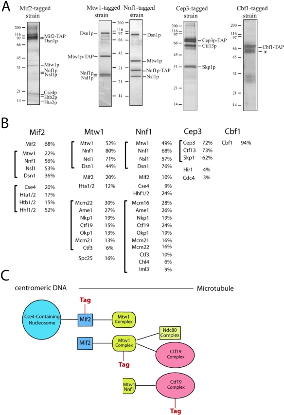
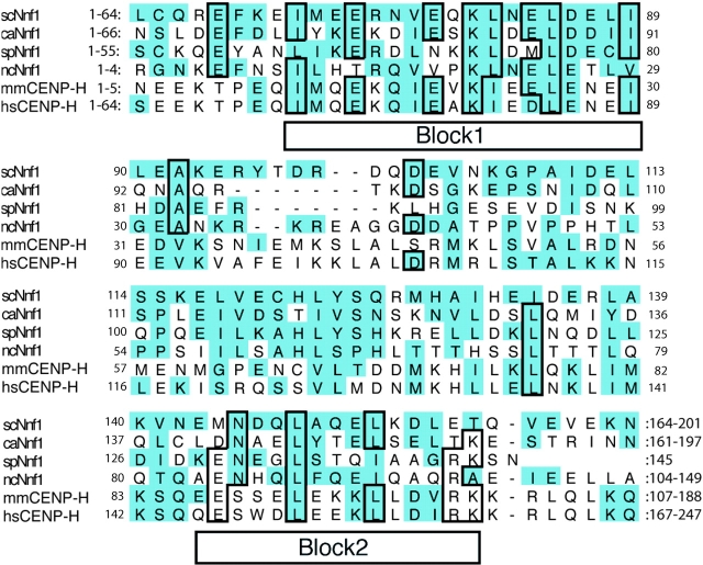
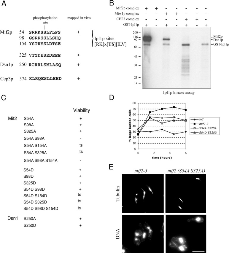
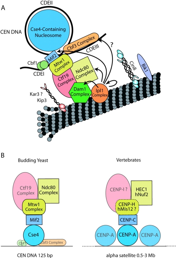
How kinetochore proteins are organized to connect chromosomes to spindle microtubules, and whether any structural and organizational themes are common to kinetochores from distantly related organisms, are key unanswered questions. Here, we used affinity chromatography and mass spectrometry to generate a map of kinetochore protein interactions. The budding yeast CENP-C homologue Mif2p specifically copurified with histones H2A, H2B, and H4, and with the histone H3-like CENP-A homologue Cse4p, strongly suggesting that Cse4p replaces histone H3 in a specialized centromeric nucleosome. A novel four-protein Mtw1 complex, the Nnf1p subunit of which has homology to the vertebrate kinetochore protein CENP-H, also copurified with Mif2p and a variety of central kinetochore proteins. We show that Mif2 is a critical in vivo target of the Aurora kinase Ipl1p. Chromatin immunoprecipitation studies demonstrated the biological relevance of these associations. We propose that a molecular core consisting of CENP-A, -C, -H, and Ndc80/HEC has been conserved from yeast to humans to link centromeres to spindle microtubules.


Figures

Figure 1.
Figure 1.
Purification of inner kinetochore proteins. (A) Silver-stained SDS-PAGE gels showing the purification of Mif2p, Cbf1p, the Mtw1 complex, and the CBF3 complex from yeast protein extracts. An asterisk denotes background bands corresponding to the heat shock proteins Ssa1 and Ssa2. (B) Percent sequence coverage obtained from mass spectrometric analysis of each purification shown in A. Brackets show proteins organized into subcomplexes. (C) Schematic diagram showing the associations between the different subcomplexes suggested by their copurification.
Figure 2.
Figure 2.
Nnf1p is homologous to vertebrate CENP-H. Sequence alignments of four fungal Nnf1p sequences and two vertebrate CENP-H sequences. The conserved blocks identified by Block Maker are indicated. Homologous residues are shaded; and identical residues are boxed (mm = Mus musculus; hs = Homo sapiens; sc = Saccharomyces cerevisiae; ca = Candida albicans; sp = Schizosaccharomyces pombe; and nc = Neuorospora crassa).
Figure 3.
Figure 3.
Analysis of kinetochore subcomplex organization by ChIP. Agarose gels (left column) showing ChIP analysis of TAP-tagged kinetochore proteins in various mutant backgrounds. CEN3 DNA was amplified by PCR from either total chromatin solution (Total, serial dilutions 1:32, 1:64, and 1:128), an immunoprecipitate (IP), or a mock-treated control (−). Control reactions show no amplification of noncentromeric DNA (PGK1) in the immunoprecipitates. Quantitation of the ChIP results (right column): the amount of CEN3 DNA in the immunoprecipitate at the permissive (25°C) and restrictive temperature (37°C) is expressed as the percentage of the DNA in the lysate. Western blotting demonstrated that there was no significant change in the level of the tagged proteins 3 h after shifting to the restrictive temperature (Fig. S3, available at http://www.jcb.org/cgi/content/full/10.1083/jcb.200305100/DC1).
Figure 4.
Figure 4.
Phosphoregulation of inner kinetochore proteins. (A) Phosphorylation sites identified in inner kinetochore proteins by mass spectrometry. (B) Mif2p and Dsn1p are direct targets of Ipl1p in vitro. Mif2, Mtw1, and CBF3 inner kinetochore complexes were purified and dephosphorylated with λ-phosphatase. In vitro phosphorylation with E. coli GST-Ipl1p was analyzed by autoradiography. Comparison with a Coomassie-stained gel (not depicted) demonstrates that Mif2p and Dsn1p are direct targets of Ipl1p. (C) Mutational analysis of the phosphorylation sites in inner kinetochore proteins. Growth on rich medium (YPD) is indicated (ts, temperature sensitive). (D) mif2 temperature-sensitive phosphorylation mutants display a metaphase arrest: cells were grown to mid-log phase and shifted to the restrictive temperature of 37°C at t = 0. The percentage of large-budded cells was determined in fixed and sonicated samples at the indicated time points. (E) Analysis of mif2 mutants 3 h after shift to 37°C. mif2-3 mutants display large-budded cells with broken down or weakened spindles (left column). The mif2 (S54A S325A) mutant shows large-budded cells with a short spindle and a single DNA mass. Bar, 5 μm.
Figure 5.
Figure 5.
Revised model of budding yeast kinetochore structure. (A) Updated model of the budding yeast kinetochore incorporating the interaction between Mif2p and the centromeric nucleosome, the positioning of the Mtw1 complex and the newly identified Ipl1p target. (B) Illustration of a structural core of conserved proteins based on the copurification and ChIP experiments. A dotted line indicates a putative complex between vertebrate CENP-H and hMis12 that awaits experimental verification.

References

    1. Cheeseman, I.M., C. Brew, M. Wolyniak, A. Desai, S. Anderson, N. Muster, J.R. Yates, T.C. Huffaker, D.G. Drubin, and G. Barnes. 2001. Implication of a novel multiprotein Dam1p complex in outer kinetochore function. J. Cell Biol. 155:1137–1146. - PMC - PubMed
    1. Cheeseman, I.M., D.G. Drubin, and G. Barnes. 2002. a. Simple centromere, complex kinetochore: linking spindle microtubules and centromeric DNA in budding yeast. J. Cell Biol. 157:199–203. - PMC - PubMed
    1. Cheeseman, I.M., S. Anderson, M. Jwa, E.M. Green, J. Kang, J.R. Yates, C.S.M. Chan, D.G. Drubin, and G. Barnes. 2002. b. Phospho-regulation of kinetochore-microtubule attachments by the Aurora kinase Ipl1p. Cell. 111:163–172. - PubMed
    1. Cleveland, D.W., Y. Mao, and K.F. Sullivan. 2003. Centromeres and kinetochores: from epigenetics to mitotic checkpoint signalling. Cell. 112:407–421. - PubMed
    1. Enquist-Newman, M., I.M. Cheeseman, D. Van Goor, D.G. Drubin, P. Meluh, and G. Barnes. 2001. Dad1p, third component of the Duo1p/Dam1p complex involved in kinetochore function and mitotic spindle integrity. Mol. Biol. Cell. 12:2601–2613. - PMC - PubMed

Publication types

MeSH terms