. 2003 Oct 27;163(2):363-74.
doi: 10.1083/jcb.200305130.

A dual-functional paramyxovirus F protein regulatory switch segment: activation and membrane fusion

Affiliations

A dual-functional paramyxovirus F protein regulatory switch segment: activation and membrane fusion

Charles J Russell et al. J Cell Biol. .

Abstract

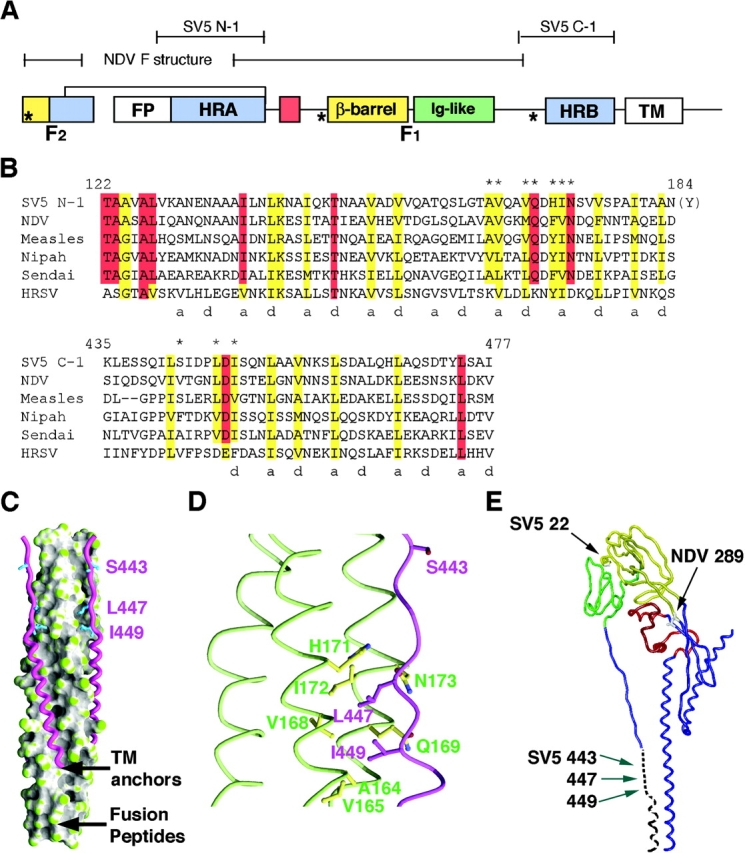
Many viral fusion-mediating glycoproteins couple alpha-helical bundle formation to membrane merger, but have different methods for fusion activation. To study paramyxovirus-mediated fusion, we mutated the SV5 fusion (F) protein at conserved residues L447 and I449, which are adjacent to heptad repeat (HR) B and bind to a prominent cavity in the HRA trimeric coiled coil in the fusogenic six-helix bundle (6HB) structure. These analyses on residues L447 and I449, both in intact F protein and in 6HB, suggest a metamorphic region around these residues with dual structural roles. Mutation of L447 and I449 to aliphatic residues destabilizes the 6HB structure and attenuates fusion activity. Mutation of L447 and I449 to aromatic residues also destabilizes the 6HB structure despite promoting hyperactive fusion, indicating that 6HB stability alone does not dictate fusogenicity. Thus, residues L447 and I449 adjacent to HRB in paramyxovirus F have distinct roles in fusion activation and 6HB formation, suggesting this region is involved in a conformational switch.


Figures

Figure 1.
Figure 1.
Schematic diagram of the paramyxovirus fusion (F) protein. The positions of the fusion peptide (FP), HRA (residues 129–184), β-barrel domain (β-barrel), Ig-like domain (Ig-like), HRB (residues 449–477), and TM are shown. The locations of the N-1 and C-1 peptides from SV5 F and the interpretable structure of NDV F are indicated. The asterisks denote SV5 residues P22 and P443, and the NDV mutant L289A, which have been implicated previously in HN-independent fusion (Ito et al., 2000; Paterson et al., 2000; Sergel et al., 2000). (B) Sequence alignment of the N-1 and C-1 peptides derived from SV5 F with the corresponding residues of F proteins from other paramyxovirus genera. HRSV, human respiratory syncytial virus. The asterisks denote the SV5 N-1 cavity residues, the C-1 cavity-binding residues L447 and I449, and residue 443, which has a hyperactive fusion phenotype when mutated to a proline residue. (C) High resolution structure of the SV5 6HB (Baker et al., 1999). The core N-1 trimer is depicted in a surface representation colored by surface curvature, and the antiparallel C-1 monomers are depicted by worm representations (magenta). (D) The packing of L447 and I449 into the hydrophobic N-1 cavity. Three N-1 (green) chains and one C-1 (magenta) chain are depicted by worm representations. (E) Worm representation of one monomer of the high resolution structure of NDV F. Arrows indicate positions of the SV5 and NDV residues implicated in HN-independent fusion (SV5 22 and 443 and NDV 289). The expected COOH-terminal continuation of the chain trace in the fusogenic/postfusion structure is depicted by the black dotted line to show where SV5 F residues 443, 447, and 449 are expected to be located. The structures in both C and E have the same orientations, with the NH2 termini of HRA at the bottoms of both panels.
Figure 2.
Figure 2.
Biophysical data on SV5 6HBs formed by N-1 and C-1 L447 and I449 mutants. (A) Thermal melts of L447 6HBs monitored by circular dichroism at 222 nm at 10 μM concentration each of N-1 and C-1 in 2 M GuHCl (PBS buffer, pH 7.0). (B) Thermal melts of I449 6HBs (same conditions as A). (C) Peptide inhibition dose–response curves measured by the luciferase assay for the L447 C-peptide mutants. Error bars repre- sent triplicate experiments. (D) Dose– response curves for the I449 C-peptide mutants. Conditions identical to C. (E) Representative analytical ultracentrifugation data (20,000 rpm) for I449F at 20°C in PBS (pH 7.0) at 10 μM concentration each of N-1 and C-1. A fit of the data to a single-ideal species model results in an observed mass of 32,470 D (calculated mass is 33,225 D) and no systematic deviation in residuals. (F) Correlation of C-1 inhibitory potency and N-1/C-1 6HB stability. C-1 peptide mutants at L447 and I449 were tested for inhibition of cell–cell fusion by the T7 luciferase reporter gene assay. The log IC50 values are plotted against the Tm of the corresponding N-1/C-1 complex. The Tm of the wt 6HB was estimated as 100°C. Error bars represent triplicate experiments.
Figure 3.
Figure 3.
Cell–cell fusion assays. (A) Representative photomicrographs of syncytia formed between BHK cells expressing the F mutants in the presence and absence of HN coexpression (20 h after transfection). Arrows point to syncytia. Bar, 200 μm. (B and C) Cell–cell fusion monitored by the luciferase reporter gene assay in the presence (B) and absence (C) of HN coexpression. The data in both panels are normalized to 100% fusion corresponding to the luciferase activity resulting from coexpression of HN and F (2 μg F DNA). F protein expression levels were determined by flow cytometry using the mAb F1a. Open boxes correspond to titrating increasing amounts of wt F DNA. Closed circles correspond to transfection of 2 μg F mutant DNA. Error bars represent triplicate experiments.
Figure 4.
Figure 4.
Cell–cell fusion monitored by dye transfer. Target cell erythrocytes (RBCs) were labeled with the green fluorescent dye 6-CF, and effector CV-1 cells coexpressing SV5 F mutants and HN were labeled with the red fluorescent dye SYTO-17. Effector CV-1 cells were infected with vaccinia virus vTF7-3 and transfected with F and HN DNA for 4 h at 37°C, incubated for 18 h at 33°C, labeled with SYTO-17 for 1 h at 37°C, coincubated with CF-labeled RBCs for 1 h at 4°C, incubated for 15 min at 37°C, and incubated on ice before confocal microscopic visualization. Membrane fusion is observed as transfer of green CF from the small RBCs to the large red-labeled CV-1 effector cells, resulting in a yellow appearance in the merge image. Large syncytium formation before 37°C incubation of effector–target cell complexes but low dye transfer by I449F and I449W or no dye transfer by L447F and L447W (denoted by arrows) is consistent with significant inactivation of the aromatic mutants by the time the complexes were incubated at 37°C. Incubation of RBC/CV-1 cell complexes for 15 min at 37°C resulted in a loss of RBC binding, consistent with the fusion deficiencies of the F proteins containing aliphatic mutations arising in an F protein intermediate preceding the prehairpin intermediate. The prehairpin intermediate retains bound RBCs during C-1 inhibition presumably due to insertion of the fusion peptide into the target membrane. Images are cropped one-quarter field views. Bar, 200 μm.
Figure 5.
Figure 5.
Cell–cell fusion measured by CF dye transfer from target RBCs to effector CV-1 cells coexpressing SV5 HN and F proteins containing mutations at L447 and I449. Conditions of the assay were similar to those detailed in Fig. 4, except either the incubation temperature (A) or incubation time (B) were varied to probe fusion kinetics. Averages and error bars are from statistical analyses of 3–5 microscopic fields. (A) Temperature dependence of fusion measured by CF dye transfer. Effector–target cell complexes were incubated at various temperatures for 5 min. No dye transfer was observed after incubation at any of the reported temperatures for no F DNA transfected or for the mutants L447F, L447W, and I449G. The F proteins containing the aliphatic mutations have increasing fusion extents as the incubation temperature is increased, whereas the F proteins containing the aromatic mutations I449F and I449W reach maximal fusion extents at a temperature as low as 22°C. (B) Kinetics of CF dye transfer for wt and the aromatic mutants at L447 and I449. In the presence of HN coexpression, wt F reached a maximal fusion extent within 5 min and F I449F and F I449W reached a maximum within 2.5 min. Cotransfection of uncleaved influenza virus Udorn HA (fusion inactive) in addition to SV5 F and HN led to retention of target RBCs after 15 min, but did not lead to an increase in the extent of dye transfer (not depicted).
Figure 6.
Figure 6.
The F proteins containing aromatic mutations cause efficient dye transfer. Cell–cell fusion measured by CF dye transfer from target RBCs to effector CV-1 cells coexpressing SV5 HN and FR3 (trypsin-inducible cleavage mutation of 5 Arg to 3 Arg residues at the cleavage site) proteins containing aromatic mutations at L447 and I449. CV-1 effector cells were incubated at 4°C for 1 h either in the presence (+T) or absence (−T) of 10 μg/ml TPCK-trypsin, subsequently coincubated with target RBCs at 4°C for 1 h and then incubated at either 22 or 37°C for 15 min. Trypsin-cleaved FR3 mutants with aromatic mutations at 447 and 449 efficiently cause CF dye transfer at both 22 and 37°C, whereas the pseudo wt FR3 mutant (with only the 5 Arg to 3 Arg mutation at the cleavage site) does not cause dye transfer at 22°C. Bar, 20 μm.
Figure 7.
Figure 7.
Model of the paramyxovirus fusion mechanism. (A) Previous reports have shown the paramyxovirus F protein adopts a series of conformations while mediating membrane fusion (for review see Russell et al., 2001); the native structure (which is in a metastable conformation), a temperature-arrested intermediate (which forms after HN binds its receptor at nonfusion-permissive temperatures and is susceptible to N-1 binding), a prehairpin intermediate (which adheres to target cells independent of HN and is susceptible to C-1 binding), and the fusogenic form of F (which couples 6HB formation to membrane merger). The present work is consistent with residues 447 and 449 having distinct interaction sites on the F protein, one in the native structure, and the other in the cavity formed by HRA trimers in the 6HB of the fusogenic form. The strong correlation between 6HB stability and C-peptide inhibitory potency in the present work and the coincidence of C-1 inhibition and F protein binding in a previous work (Russell et al., 2001) show that C-1 inhibits membrane fusion by binding to transiently exposed HRA triple-stranded coiled coils in the prehairpin intermediate. (B) Biophysical experiments on the N-1/C-peptide mutants show that the aliphatic (al) and aromatic (ar) mutations at L447 and I449 decrease the amount of energy released by mutant 6HB formation, whereas functional experiments on the F mutants are consistent with the aromatic (ar) mutations decreasing and the aliphatic (al) mutations increasing the activation energy of the native state. The hyperactive fusion phenotype of the aromatic mutants is consistent with the facile activation (and subsequent inactivation in the absence of a fusion target) of these F mutants overcompensating for the decrease in energy released by mutant 6HB formation.

References

    1. Armstrong, R.T., A.S. Kushnir, and J.M. White. 2000. The transmembrane domain of influenza hemagglutinin exhibits a stringent length requirement to support the hemifusion to fusion transition. J. Cell Biol. 151:425–437. - PMC - PubMed
    1. Baker, K.A., R.E. Dutch, R.A. Lamb, and T.S. Jardetzky. 1999. Structural basis for paramyxovirus-mediated membrane fusion. Mol. Cell. 3:309–319. - PubMed
    1. Buckland, R., E. Malvoisin, P. Beauverger, and F. Wild. 1992. A leucine zipper structure present in the measles virus fusion protein is not required for its tetramerization but is essential for fusion. J. Gen. Virol. 73:1703–1707. - PubMed
    1. Bullough, P.A., F.M. Hughson, J.J. Skehel, and D.C. Wiley. 1994. Structure of influenza haemagglutinin at the pH of membrane fusion. Nature. 371:37–43. - PubMed
    1. Calder, L.J., L. Gonzalez-Reyes, B. Garcia-Barreno, S.A. Wharton, J.J. Skehel, D.C. Wiley, and J.A. Melero. 2000. Electron microscopy of the human respiratory syncytial virus fusion protein and complexes that it forms with monoclonal antibodies. Virology. 271:122–131. - PubMed

Publication types

MeSH terms