. 2003 Nov;77(22):12022-32.
doi: 10.1128/jvi.77.22.12022-12032.2003.

DC-SIGN and L-SIGN can act as attachment receptors for alphaviruses and distinguish between mosquito cell- and mammalian cell-derived viruses

Affiliations

DC-SIGN and L-SIGN can act as attachment receptors for alphaviruses and distinguish between mosquito cell- and mammalian cell-derived viruses

William B Klimstra et al. J Virol. 2003 Nov.

Erratum in

  • J Virol. 2004 Jul;78(14):7862

Abstract

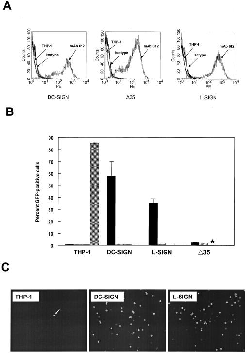
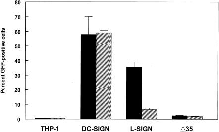
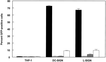
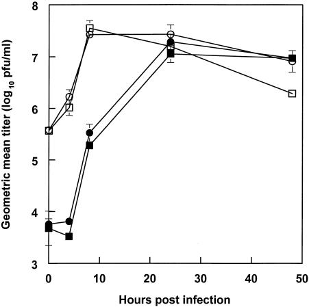
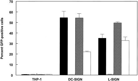
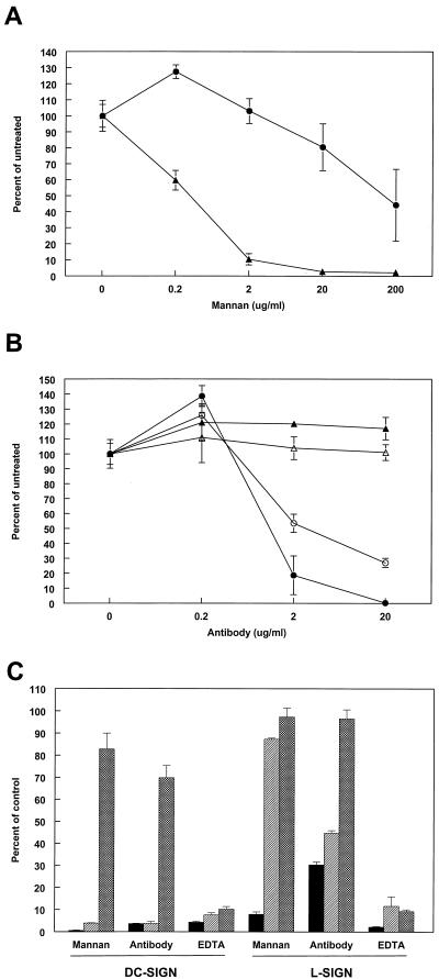
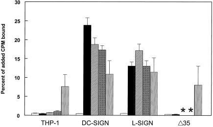
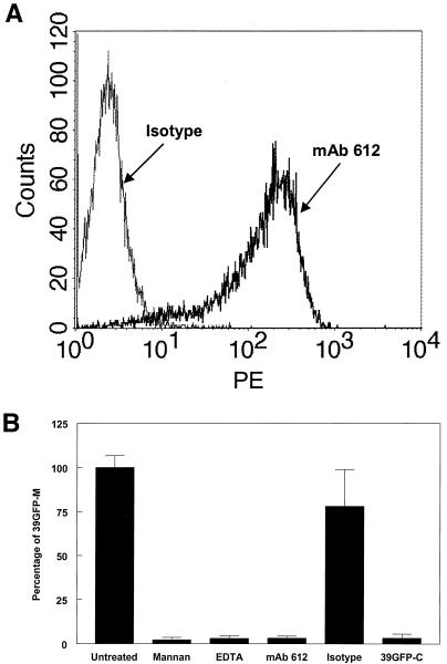
C-type lectins such as DC-SIGN and L-SIGN, which bind mannose-enriched carbohydrate modifications of host and pathogen proteins, have been shown to bind glycoproteins of several viruses and facilitate either cis or trans infection. DC-SIGN and L-SIGN are expressed in several early targets of arbovirus infection, including dendritic cells (DCs) and cells of the reticuloendothelial system. In the present study, we show that DC-SIGN and L-SIGN can function as attachment receptors for Sindbis (SB) virus, an arbovirus of the Alphavirus genus. Human monocytic THP-1 cells stably transfected with DC-SIGN or L-SIGN were permissive for SB virus replication, while untransfected controls were essentially nonpermissive. The majority of control THP-1 cells were permissive when attachment and entry steps were eliminated through electroporation of virus transcripts. Infectivity for the DC-SIGN/L-SIGN-expressing cells was largely blocked by yeast mannan, EDTA, or a DC-SIGN/L-SIGN-specific monoclonal antibody. Infection of primary human DCs by SB virus was also dependent upon SIGN expression by similar criteria. Furthermore, production of virus particles in either C6/36 mosquito cells or CHO mammalian cells under conditions that limited complex carbohydrate content greatly increased SB virus binding to and infection of THP-1 cells expressing these lectins. C6/36-derived virus also was much more infectious for primary human DCs than CHO-derived virus. These results suggest that (i) lectin molecules such as DC-SIGN and L-SIGN may represent common attachment receptor molecules for arthropod-borne viruses, (ii) arbovirus particles produced in and delivered by arthropod vectors may preferentially target vertebrate host cells bearing these or similar lectin molecules, and (iii) a cell line has been identified that can productively replicate alphaviruses but is deficient in attachment receptors.


Figures

FIG. 1.
FIG. 1.
DC-SIGN/L-SIGN expression and SB virus infection of THP-1 cells. (A) Histogram of flow cytometric analysis of cell surface DC-SIGN/L-SIGN expression on THP-1 control and transfected cells. Control THP-1 cells were stained with MAb 612, and transfected cells were stained with MAb 612 or the isotype control MAb. (B) Infectivity of 39GFP-M (solid bars), 39GFP-C (hatched bars), or CHOK1-derived HS binding mutant (S1K70GFP) SB virus (open bars) for control or transfected THP-1 cells and electroporation efficiency of TR339GFP transcripts (crosshatched bars) for control THP-1 cells. *, S1K70 infectivity not measured. (C) Fluorescence micrographs (magnification, ×100) of THP-1 cells infected with 39GFP-M virus. Equal numbers of each THP-1 cell type were infected with 39GFP-M as described in Materials and Methods and then diluted 10-fold into 96-well plates, incubated for 12 h, and photographed on the fluorescence microscope. Bright-field observations confirmed that each view shown contained similar numbers of cells. The arrow indicates a rare GFP-positive cell in control THP-1 cells.
FIG. 2.
FIG. 2.
Effect of infection temperature on infectivity of 39GFP-M for THP-1 cells. Solid bars, 37°C; hatched bars, 4°C.
FIG. 3.
FIG. 3.
Infectivity of 39GFP-M (solid bars), 39-GFP-C (hatched bars), or 39GFP derived from THP-1 (crosshatched bars) or DF-1 (open bars) cells for control THP-1 or DC-SIGN/L-SIGN-expressing cells.
FIG. 4.
FIG. 4.
Growth of TR339 in cultures of transfected THP-1 cells. DC-SIGN-transfected cells (squares) or L-SIGN-transfected cells (circles) were infected with 39-M (open symbols) or 39-C (solid symbols) virus.
FIG. 5.
FIG. 5.
Infectivity of 39GFP-M (solid bars), 39GFP-C (hatched bars), 39GFP-L (crosshatched bars), or 39GFP-DMJ (open bars) virus for control THP-1 or DC-SIGN/L-SIGN-expressing cells.
FIG. 6.
FIG. 6.
Infectivity of 39GFP for cells in the presence of DC-SIGN/L-SIGN competitors or EDTA. (A) Infectivity of 39GFP-M virus for THP-1 cells expressing DC-SIGN (solid triangles) or L-SIGN (solid circles) in the presence of increasing concentrations of mannan. (B) Infectivity of 39GFP-M virus for THP-1 cells expressing DC-SIGN (solid symbols) or L-SIGN (open symbols) in the presence of increasing concentrations of MAb 612 (circles) or an isotype-matched control MAb (triangles). (C) Infectivity of 39GFP-M (solid bars), 39GFP-L (hatched bars), or 39GFP-DMJ (crosshatched bars) virus for THP-1 cells in the presence of 200 μg of mannan per ml, 20 μg of MAb 612 per ml, or 5 mM EDTA. The control for mannan- or EDTA-treated cells was untreated cells. The control for MAb 612 was 20 μg of isotype-matched MAb per ml.
FIG. 7.
FIG. 7.
Binding of radiolabeled TR339 or S1K70 virus to THP-1 control and DC-SIGN/L-SIGN-transfected cells. The viruses used were 39-C (open bars), 39-M (solid bars), 39-L (hatched bars), or 39-DMJ (crosshatched bars) and S1K70 (striped bars), which was derived from CHO cells. *, binding of TR339 derived from Lec1- and 1-DMJ-treated Pro5 CHO cells not tested on Δ35 THP-1 cells.
FIG. 8.
FIG. 8.
Flow cytometric analysis of cell surface DC-SIGN expression and infectivity of 39GFP viruses for primary human DCs. (A) Histogram showing MAb and isotype control antibody reactivity with primary human DCs. (B) Relative infectivity of 39GFP-M and 39GFP-C for human DCs and relative blocking of DC infection by 39GFP-M by 200 μg of mannan per ml, 5 mM EDTA, 20 μg of MAb 612 or isotype control antibody per ml. Approximately 30% of the cells infected with 39GFP-M were positive for GFP at 6 hpi.

References

    1. Bashirova, A. A., T. B. Geijtenbeek, G. C. van Duijnhoven, S. J. Van Vliet, J. B. Eilering, M. P. Martin, L. Wu, T. D. Martin, N. Viebig, P. A. Knolle, V. N. KewalRamani, Y. van Kooyk, and M. Carrington. 2001. A dendritic cell-specific intercellular adhesion molecule 3-grabbing nonintegrin (DC-SIGN)-related protein is highly expressed on human liver sinusoidal endothelial cells and promotes HIV-1 infection. J. Exp. Med. 193:671-678. - PMC - PubMed
    1. Bernard, K. A., W. B. Klimstra, and R. E. Johnston. 2000. Mutations in the E2 glycoprotein of Venezuelan equine encephalitis virus confer heparan sulfate interaction, low morbidity, and rapid clearance from blood of mice. Virology 276:93-103. - PubMed
    1. Byrnes, A. P., and D. E. Griffin. 1998. Binding of Sindbis virus to cell surface heparan sulfate. J. Virol. 72:7349-7356. - PMC - PubMed
    1. Chen, J.-P., J. H. Strauss, E. G. Strauss, and T. K. Frey. 1996. Characterization of the rubella virus nonstructural protease domain and its cleavage site. J. Virol. 70:4707-4713. - PMC - PubMed
    1. Curtis, B. M., S. Scharnowske, and A. J. Watson. 1992. Sequence and expression of a membrane-associated C-type lectin that exhibits CD4-independent binding of human immunodeficiency virus envelope glycoprotein gp120. Proc. Natl. Acad. Sci. USA 89:8356-8360. - PMC - PubMed

Publication types

LinkOut - more resources