. 2003 Nov;77(22):12140-51.
doi: 10.1128/jvi.77.22.12140-12151.2003.

Human immunodeficiency virus type 1 enters primary human brain microvascular endothelial cells by a mechanism involving cell surface proteoglycans independent of lipid rafts

Affiliations

Human immunodeficiency virus type 1 enters primary human brain microvascular endothelial cells by a mechanism involving cell surface proteoglycans independent of lipid rafts

Elias G Argyris et al. J Virol. 2003 Nov.

Abstract

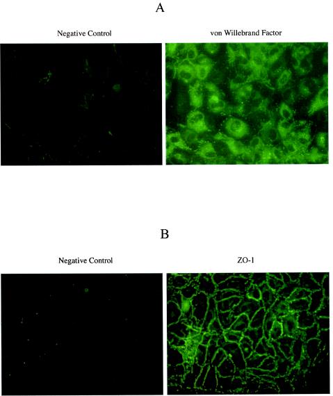
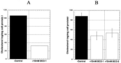
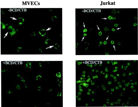
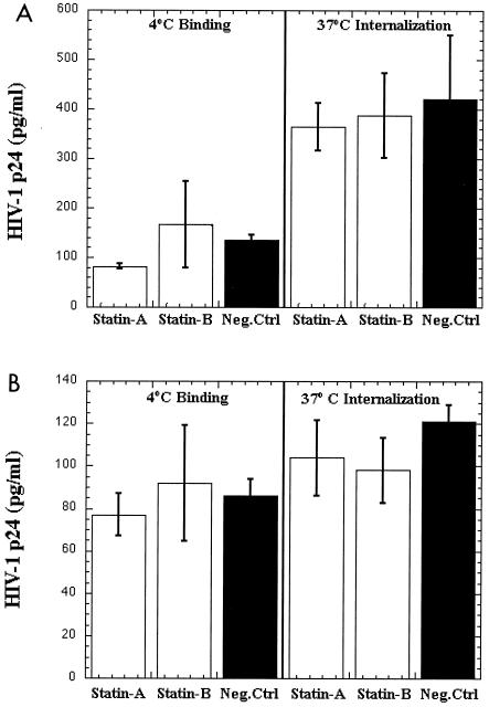
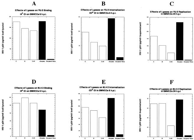
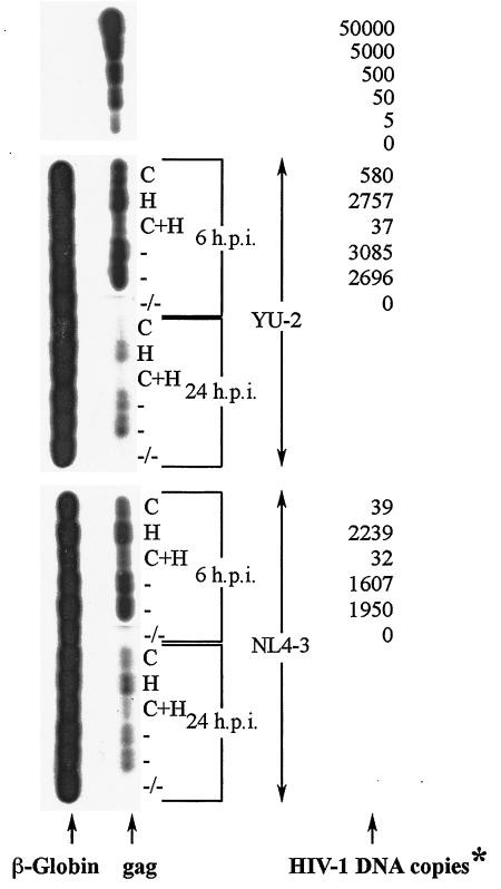
Several studies have reported a crucial role for cholesterol-enriched membrane lipid rafts and cell-associated heparan sulfate proteoglycans (HSPGs), a class of molecules that can localize in lipid rafts, in the entry of human immunodeficiency virus type 1 (HIV-1) into permissive cells. For the present study, we examined the role of these cell surface moieties in HIV-1 entry into primary human brain microvascular endothelial cells (BMVECs), which represent an important HIV-1 central nervous system-based cell reservoir and a portal for neuroinvasion. Cellular cholesterol was depleted by exposure to beta-cyclodextrins and 3-hydroxy-3-methylglutaryl (HMG)-coenzyme A reductase inhibitors (statins), the loss of cholesterol was quantitated, and disruption of membrane rafts was verified by immunofluorescence. Nevertheless, these treatments did not affect binding of several strains of HIV-1 virions to BMVECs at 4 degrees C or their infectivities at 37 degrees C. In contrast, we confirmed that cholesterol depletion and raft disruption strongly inhibited HIV-1 binding and infection of Jurkat T cells. Enzymatic digestion of cell-associated HSPGs on human BMVECs dramatically inhibited HIV-1 infection, and our data from quantitative HIV-1 DNA PCR analysis strongly suggest that cell-associated chondroitin sulfate proteoglycans greatly facilitate infective entry of HIV-1 into human BMVECs. These findings, in combination with our earlier work showing that human BMVECs lack CD4, indicate that the molecular mechanisms for HIV-1 entry into BMVECs are fundamentally different from that of viral entry into T cells, in which lipid rafts, CD4, and probably HSPGs play important roles.


Figures

FIG. 1.
FIG. 1.
Immunofluorescence microscopy of primary isolated human BMVECs for endothelial cell markers von Willebrand factor (A) and ZO-1 (B). Primary isolated human BMVECs were cultured on 2-well chamber slides for 48 h. The cells were fixed with 3.5% formaldehyde and incubated with primary antibodies against the respective cell markers. The negative control represents immunostaining of primary human BMVECS with secondary antibody. The figure is representative of 10 independent studies.
FIG. 2.
FIG. 2.
Depletion of cellular cholesterol by BCDs in primary human BMVECs (A) and Jurkat T cells (B). Cellular cholesterol was measured by cholesterol oxidase-based colorimetric assay as described in Materials and Methods. The cholesterol content of cells was normalized to total cellular protein. Shown are the results of an experiment with primary human BMVECs and of two independent studies with the control Jurkat T cells. BCD-1, cells pretreated with 10 mM randomly methylated BCD (TRMBP); BCD-2, cells pretreated with 10 mM hydroxypropyl BCD (THPBP); −Control, untreated cells.
FIG. 3.
FIG. 3.
Staining of primary human BMVECs and Jurkat T cells for GM1-lipid rafts. Cells were first fixed with 3.5% formaldehyde on 2-well chamber slides and then incubated with 2 μg of fluorescent conjugate (Alexa Fluor 488-CTB) per ml for 2 h at room temperature. The figure is representative of over four independent studies. −BCD/CTB, cells not treated with BCDs and stained with Alexa-CTB against GM1, demonstrating a distinct ring pattern (white arrows); +BCD/CTB, cells treated with BCDs and stained with Alexa-CTB against GM1.
FIG. 4.
FIG. 4.
Effects of BCDs on HIV-1 binding and entry into primary human BMVECs. (A) Effects of different BCD compounds on HIV-1 CCR-5-tropic YU-2 binding (4°C) and entry (37°C) into primary human BMVECs. (B) Effects of BCDs on HIV-1 X4-tropic NL4-3 binding (4°C) and entry (37°C) into BMVECs. (C) Effects of BCDs on HIV-1 NL4-3 Env Vpr binding (4°C) and entry (37°C) into BMVECs. The graphs represent the mean values of two independent experiments. BCD-1, cells pretreated with 10 mM randomly methylated BCD (TRMBP); BCD-2, cells pretreated with 10 mM hydroxypropyl BCD (THPBP); Neg.Ctrl, untreated BMVECs.
FIG. 5.
FIG. 5.
Effects of statins on HIV-1 binding and entry into primary human BMVECs. (A) Effects of different statins on HIV-1 CCR-5-tropic YU-2 binding (4°C) and entry (37°C) into primary human BMVECs. (B) Effects of different statins on HIV-1 X4-tropic NL4-3 binding (4°C) and entry (37°C) into BMVECs. The graphs represent the mean values of two independent experiments. Statin-A, cells pretreated with 3 μM atorvastatin; Statin-B, cells pretreated with 3 μM pravastatin; Neg.Ctrl, untreated BMVECs.
FIG. 6.
FIG. 6.
Effects of BCDs and statins on HIV-1 binding and entry into Jurkat T cells. (A) Effects of hydroxypropyl BCD and/or mevinolin on HIV-1 NL4-3 binding (4°C) on Jurkat T cells. (B) Effects of hydroxypropyl BCD and/or mevinolin on HIV-1 NL4-3 entry (37°C) into Jurkat T cells. The graphs represent the mean values of two independent experiments. THPBP, 10 mM hydroxypropyl BCD; Neg.Ctrl, untreated Jurkat T cells.
FIG. 7.
FIG. 7.
Immunofluorescence microscopy of primary isolated human BMVECs for heparan and chondroitin sulfate proteoglycans. (A) The negative control represents immunostaining of primary human BMVECs with secondary antibody. (B) Primary human BMVECs stained for cell surface heparan sulfate moieties. (C) Immunostaining of primary human BMVECs pretreated with 5 U of heparitinase confirms the removal of cell surface heparan sulfate moieties. (D) Primary human BMVECs stained for cell surface chondroitin sulfate moieties. (E) Immunostaining of primary human BMVECs pretreated with 5 U of chondroitinase-ABC confirms the removal of cell surface chondroitin sulfate moieties. The figure is representative of three independent studies.
FIG. 8.
FIG. 8.
Effects of lyases (heparitinase and chondroitinase) on HIV-1 binding, entry, and replication in primary human BMVECs. Effects of lyases on HIV-1 YU-2 binding (4°C) (A), internalization (37°C) (B), and replication (C) in BMVECs and HIV-1 NL4-3 binding (4°C) (D), internalization (37°C) (E), and replication (F) in BMVECs. Cells were treated according to standard procedures, as described in Materials and Methods. The graphs represent a single experiment (samples were analyzed in duplicate). C, BMVECs pretreated with 5 U of chondroitinase per ml; H, BMVECs pretreated with 5 U of heparitinase per ml; C+H, BMVECs pretreated with a combination of 5 U of chondroitinase per ml and 5 U of heparitinase per ml; −Enzyme, untreated BMVECs; −Enzyme/−Virus, untreated and uninfected BMVECs.
FIG. 9.
FIG. 9.
Detection of proviral DNA (YU-2 and NL4-3) at 6 and 24 h postinfection in primary human BMVECs pretreated with lyases or left untreated. Cells were treated according to standard procedures, as described in Materials and Methods. Cellular DNA was extracted from BMVECs and amplified by PCR with gag SK38 and SK39 as the primer pair. As a means of confirming that each sample contained similar quantities of cellular DNA before PCR, β-globin DNA (left) was also amplified in each sample, with PCO3 and PCO4 as the primer pair. The HIV-1 DNA standards (50,000 to 0 copies; top) were prepared from ACH-2 cells (one proviral copy per cell) and amplified by PCR at the same time as all the samples; values (right) for each sample are given as numbers of copies per 2.5 × 10⁵ cells and correspond to the matching HIV-1 gag DNA bands (left). *, at 24 h postinfection, samples were analyzed by detection only, not by DNA-copy quantification (between 1 and 5 copies); h.p.i., hours postinfection; C, BMVECs pretreated with 5 U of chondroitinase per ml; H, BMVECs pretreated with 5 U of heparitinase per ml; C+H, BMVECs pretreated with a combination of 5 U of chondroitinase per ml and 5 U of heparitinase per ml; −, untreated BMVECs; −/−, untreated and uninfected BMVECs.

References

    1. Allain, C. C., L. S. Poon, C. S. Chan, W. Richmond, and P. C. Fu. 1974. Enzymatic determination of total serum cholesterol. Clin. Chem. 20:470-475. - PubMed
    1. An, S. F., M. Groves, F. Gray, and F. Scaravilli. 1999. Early entry and widespread cellular involvement of HIV-1 DNA in brains of HIV-1 positive asymptomatic individuals. J. Neuropathol. Exp. Neurol. 58:1156-1162. - PubMed
    1. Anderson, R. G., and K. Jacobson. 2002. A role for lipid shells in targeting proteins to caveolae, rafts, and other lipid domains. Science 296:1821-1825. - PubMed
    1. Bagasra, O., E. Lavi, L. Bobroski, K. Khalili, J. P. Pestaner, R. Tawadros, and R. J. Pomerantz. 1996. Cellular reservoirs of HIV-1 in the central nervous system of infected individuals: identification by the combination of in situ polymerase chain reaction and immunohistochemistry. AIDS 10:573-585. - PubMed
    1. Bell, J. E., A. Busuttil, J. W. Ironside, S. Rebus, Y. K. Donaldson, P. Simmonds, and J. F. Peutherer. 1993. Human immunodeficiency virus and the brain: investigation of virus load and neuropathologic changes in pre-AIDS subjects. J. Infect. Dis. 168:818-824. - PubMed

Publication types