. 2003 Nov;77(22):12193-202.
doi: 10.1128/jvi.77.22.12193-12202.2003.

Engineered retargeting of viral RNA replication complexes to an alternative intracellular membrane

Affiliations

Engineered retargeting of viral RNA replication complexes to an alternative intracellular membrane

David J Miller et al. J Virol. 2003 Nov.

Abstract

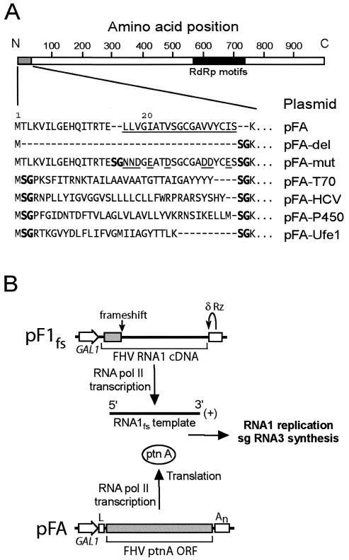
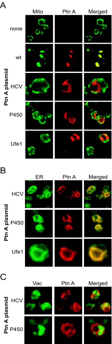
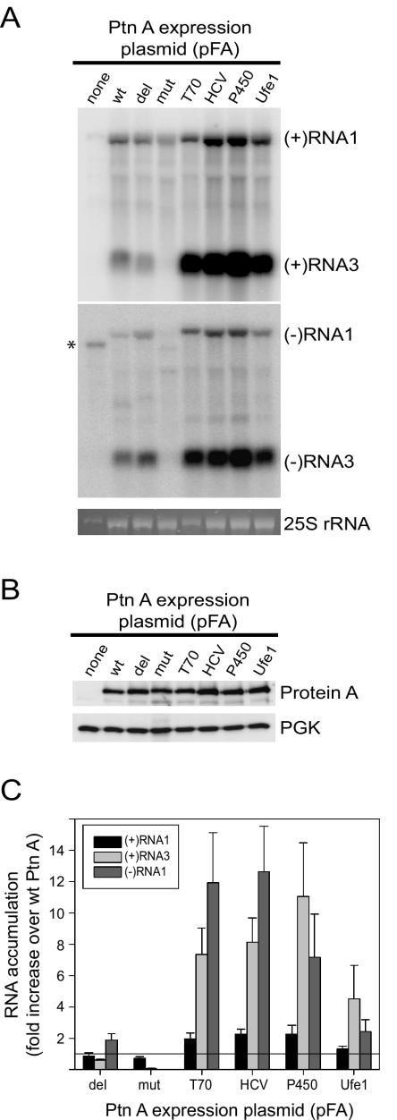
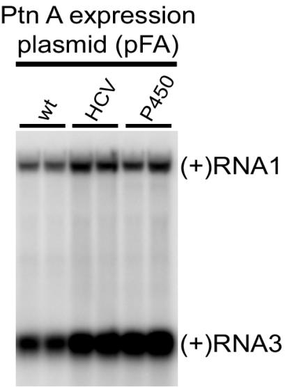
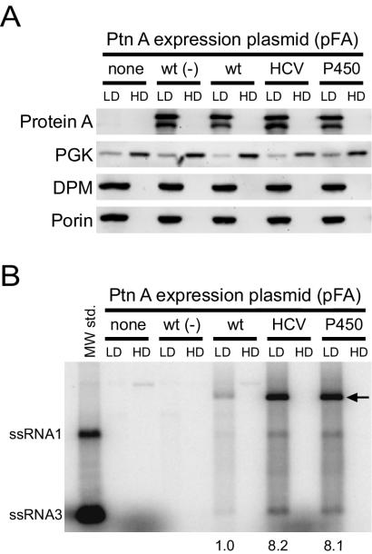
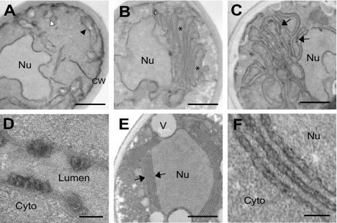
Positive-strand RNA virus replication complexes are universally associated with intracellular membranes, although different viruses use membranes derived from diverse and sometimes multiple organelles. We investigated whether unique intracellular membranes are required for viral RNA replication complex formation and function in yeast by retargeting protein A, the Flock House virus (FHV) RNA-dependent RNA polymerase. Protein A, the only viral protein required for FHV RNA replication, targets and anchors replication complexes to outer mitochondrial membranes in part via an N-proximal sequence that contains a transmembrane domain. We replaced the FHV protein A mitochondrial outer membrane-targeting sequence with the N-terminal endoplasmic reticulum (ER)-targeting sequence from the yeast NADP cytochrome P450 oxidoreductase or inverted C-terminal ER-targeting sequences from the hepatitis C virus NS5B polymerase or the yeast t-SNARE Ufe1p. Confocal immunofluorescence microscopy confirmed that protein A chimeras retargeted to the ER. FHV subgenomic and genomic RNA accumulation in yeast expressing ER-targeted protein A increased 2- to 13-fold over that in yeast expressing wild-type protein A, despite similar protein A levels. Density gradient flotation assays demonstrated that ER-targeted protein A remained membrane associated, and in vitro RNA-dependent RNA polymerase assays demonstrated an eightfold increase in the in vitro RNA synthesis activity of the ER-targeted FHV RNA replication complexes. Electron microscopy showed a change in the intracellular membrane alterations from a clustered mitochondrial distribution with wild-type protein A to the formation of perinuclear layers with ER-targeted protein A. We conclude that specific intracellular membranes are not required for FHV RNA replication complex formation and function.


Figures

FIG. 1.
FIG. 1.
(A) Chimeric FHV protein A sequences and plasmid designations. A schematic of protein A with the viral RdRp motif region represented by the solid bar is shown on top for reference. The core predicted protein A TMD from Leu 17 to Ser 35 is underlined (32). The hydrophobic-to-hydrophilic amino acid mutations in pFA-mut are also underlined. The unique BspEI site used to generate the chimeric protein A constructs introduced a Ser-Gly at the insertion sequence junctions (shown in boldface). The dashes indicate no amino acids and are included for alignment purposes. (B) Schematic of plasmid-directed FHV RNA replication in yeast. RNA1 templates with authentic viral 5′ and 3′ termini are generated from pF1fs through precise placement of the GAL1 promoter start site and a hepatitis δ ribozyme (Rz), respectively, and the frameshift at the indicated location disrupts translation. The GAL1 leader (L) and CYC1 polyadenylation signal (An) flanking the protein A open reading frame (ORF) in pFA or derivatives thereof disrupt its activity as a viral RNA replication template but enhance its RNA polymerase II-directed transcription and translation. pFA-derived protein A (ptnA) subsequently directs RNA1 replication and subgenomic (sg) RNA3 synthesis from an RNA1 template transcribed from pF1fs.
FIG. 2.
FIG. 2.
Chimeric protein A is retargeted to the ER. (A) Yeasts expressing pF1fs plus a control plasmid lacking the protein A open reading frame (top row), pFA (second row), pFA-HCV (third row), pFA-P450 (fourth row), or pFA-Ufe1 (bottom row) were immunostained with rabbit anti-protein A (Ptn A) and mouse anti-CoxIII for mitochondria (mito), followed by Texas red-labeled goat anti-rabbit and fluorescein isothiocyanate-labeled goat anti-mouse secondary antibodies. Representative images for CoxIII (left; green), protein A (middle; red), and merged signals (right) are shown. The merged images represent a digital superimposition of red and green signals in which areas of fluorescence colocalization are yellow. The images for pFA-HCV (third row) are shown at approximately twice the magnification of the other immunofluorescence images. (B) Yeast cells expressing pF1fs plus pFA-HCV (top row), pFA-P450 (middle row), or pFA-Ufe1 (bottom row) were immunostained with rabbit anti-protein A and Oregon Green 488-labeled concanavalin A, followed by Texas red-labeled goat anti-rabbit secondary antibodies. Representative confocal images for conconavalin A (left; green), protein A (middle; red), and merged signals (right) are shown. The images for pFA-Ufe1 (bottom row) are shown at approximately twice the magnification of the other immunofluorescence images. Concanavalin A is a lectin that selectively binds α-mannopyranosyl and α-glucopyranosyl residues. Initial experiments demonstrated that the fluorescence pattern of concanavalin A reactivity in yeast colocalized with the immunofluorescence pattern of Kar2p, a yeast ER-resident protein (data not shown). (C) Yeast cells expressing pF1fs plus pFA-HCV (top row) or pFA-P450 (bottom row) were immunostained with rabbit anti-protein A and mouse anti-vacuolar ATPase for vacuoles (Vac), followed by Texas red-labeled goat anti-rabbit and fluorescein isothiocyanate-labeled goat anti-mouse secondary antibodies. Representative confocal images for vacuolar ATPase (left; green), protein A (middle; red), and merged signals (right) are shown. The images for pFA-P450 (bottom row) are shown at approximately twice the magnification of the other immunofluorescence images.
FIG. 3.
FIG. 3.
ER-targeted protein A chimeras increase FHV RNA accumulation in vivo. (A) Viral RNA accumulation in yeasts expressing pF1fs plus the indicated protein A (Ptn A) expression plasmids. The lane labels correspond to the plasmid designations shown in Fig. 1A. Total RNAs from equivalent numbers of cells were separated by electrophoresis and blotted with 32P-labeled complementary riboprobes that corresponded to nucleotides 2718 to 3064 from FHV RNA1 (38). Note that RNA3 is colinear with the 3′ end of RNA1 (13, 17) and that the probe sequence is present in both RNAs. Control Northern blots comparing known amounts of RNA1 and RNA3 in vitro transcripts showed that, with the probes and the transfer and hybridization conditions used, there was no significant difference between the detection efficiencies of RNA1 and RNA3. Thus, the band intensities reflect the molar ratios of RNA1 and RNA3. The riboprobes were either in the antisense or sense orientation and detected (+)RNA1 and (+)RNA3 (upper blot) or (−)RNA1 and (−)RNA3 (lower blot), respectively. The positions of RNA1 and RNA3 are shown on the right. The ethidium bromide-stained band of 25S rRNA is shown below the blots as a loading control. The asterisk indicates the position of a background band seen prominently in yeast expressing template only but also present to a lesser extent in all samples. This band was not present in total RNA preparations from FHV-infected Drosophila cells, whereas the (−)RNA1-labeled band was present (not shown). Subgenomic (−)RNA3 is produced in yeast replicating FHV RNA and in FHV-infected Drosophila cells and may function as a template for subgenomic (+)RNA3 synthesis (27, 38). (B) FHV protein A accumulation in yeast expressing pF1fs plus protein A expression plasmids. Total protein from equivalent numbers of cells was separated by electrophoresis and blotted with rabbit anti-protein A antiserum. The anti-PGK immunoblot is shown as a loading control. (C) Quantitative analysis of (+)RNA1, (+)RNA3, and (−)RNA1 accumulation. Northern blots were quantitated by phosphorimager analysis, and the results are expressed as the increase (n-fold) over yeast expressing pF1fs plus pFA. The horizontal line at 1 is placed for reference to the wt control. Averages and standard errors of the mean of at least three independent experiments are shown.
FIG. 4.
FIG. 4.
ER-targeted protein A (Ptn A) chimeras maintain increased FHV RNA accumulation in vivo after 72 h of induction. Total RNA from equivalent numbers of cells was separated by electrophoresis and blotted with a 32P-labeled complementary riboprobe that detected (+)RNA1 and (+)RNA3. Duplicate representative samples from yeast cells expressing pF1fs plus pFA, pFA-HCV, or pFA-P450 are shown.
FIG. 5.
FIG. 5.
ER-targeted protein A (Ptn A) chimeras are membrane-associated and have enhanced in vitro FHV RdRp activities. (A) Equilibrium density gradient fractionation of lysates from yeast cells expressing pF1fs alone (none), pFA alone [wt (−)], or pF1fs plus pFA (wt), pFA-HCV, or pFA-P450. Equal amounts of total protein from both LD and HD fractions were separated by sodium dodecyl sulfate-polyacrylamide gel electrophoresis and immunoblotted with mouse MAbs to protein A, PGK, DPM, or porin. Loading equal amounts of total protein resulted in an overrepresentation of individual proteins in the LD fraction relative to the HD fraction, thus exaggerating the residual PGK signal in the LD fraction. Protein A appears as a doublet in immunoblots of equilibrium gradient fractions, where the lower band represents a C-terminal degradation product (32). (B) In vitro RdRp assay of equilibrium density gradient fractions. Equal amounts of total proteins from both LD and HD fractions were incubated with [32P]UTP and unlabeled ribonucleotides, and the reaction products were separated by nondenaturing agarose gel electrophoresis. The positions of in vitro-transcribed ssRNA1 and ssRNA3 are shown on the left. The major reaction products corresponding to ssRNA1, ssRNA3, and presumed replicative intermediate dsRNA1 (arrow) were quantitated by phosphorimager analysis in three independent experiments, and the numbers represent the increases (n-fold) in total radiolabeled products relative to the LD fraction of yeast expressing pF1fs plus pFA.
FIG. 6.
FIG. 6.
FHV protein A expression and RNA replication induces distinct ultrastructural membrane alterations in yeast. (A) Electron micrograph of yeast expressing pF1fs only, showing normal nucleus (Nu), cell wall (CW), and mitochondria seen in longitudinal (black arrowhead) and transverse (white arrowhead) sections. (B) Electron micrograph of yeast expressing pFA only, showing clustering of membrane-bounded organelles (asterisks). (C) Electron micrograph of yeast expressing pF1fs plus pFA, showing clustered membrane-bounded organelles plus electron-dense structures (arrows). (D) Electron micrograph (higher magnification) of membrane-bounded structures projecting into the organelle lumen of yeast expressing pF1fs plus pFA. Cyto, cytoplasm. (E) Electron micrograph of yeast expressing pF1fs plus pFA-HCV, showing perinuclear membrane layers (arrows). V, vacuole. (F) Electron micrograph (higher magnification) of perinuclear membrane layers in yeast expressing pF1fs plus pFA-HCV, showing an irregular appearance. Bars = 500 (A, B, C, and E), 100 (D), and 150 (F) nm.

References

    1. Ahlquist, P., A. O. Noueiry, W. Lee, D. B. Kushner, and B. T. Dye. 2003. Host factors in positive-strand RNA virus genome replication. J. Virol. 77:8181-8186. - PMC - PubMed
    1. Ahola, T., A. Lampio, P. Auvinen, and L. Kaariainen. 1999. Semliki Forest virus mRNA capping enzyme requires association with anionic membrane phospholipids for activity. EMBO J. 18:3164-3172. - PMC - PubMed
    1. Ball, L. A. 1995. Requirements for the self-directed replication of Flock House virus RNA 1. J. Virol. 69:720-727. - PMC - PubMed
    1. Ball, L. A., and K. L. Johnson. 1998. Nodaviruses of insects, p. 225-267. In L. K. Miller and L. A. Ball (ed.), The insect viruses. Plenum Publishing Corp., New York, N.Y.
    1. Ball, L. A., and Y. Li. 1993. cis-acting requirements for the replication of Flock House virus RNA 2. J. Virol. 67:3544-3551. - PMC - PubMed

Publication types

LinkOut - more resources