. 2003 Nov 3;198(9):1427-37.
doi: 10.1084/jem.20030802. Epub 2003 Oct 27.

The balance between Pax5 and Id2 activities is the key to AID gene expression

Affiliations

The balance between Pax5 and Id2 activities is the key to AID gene expression

Hiroyuki Gonda et al. J Exp Med. .

Abstract

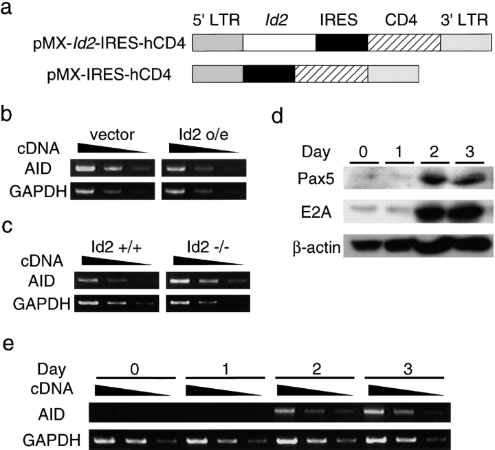
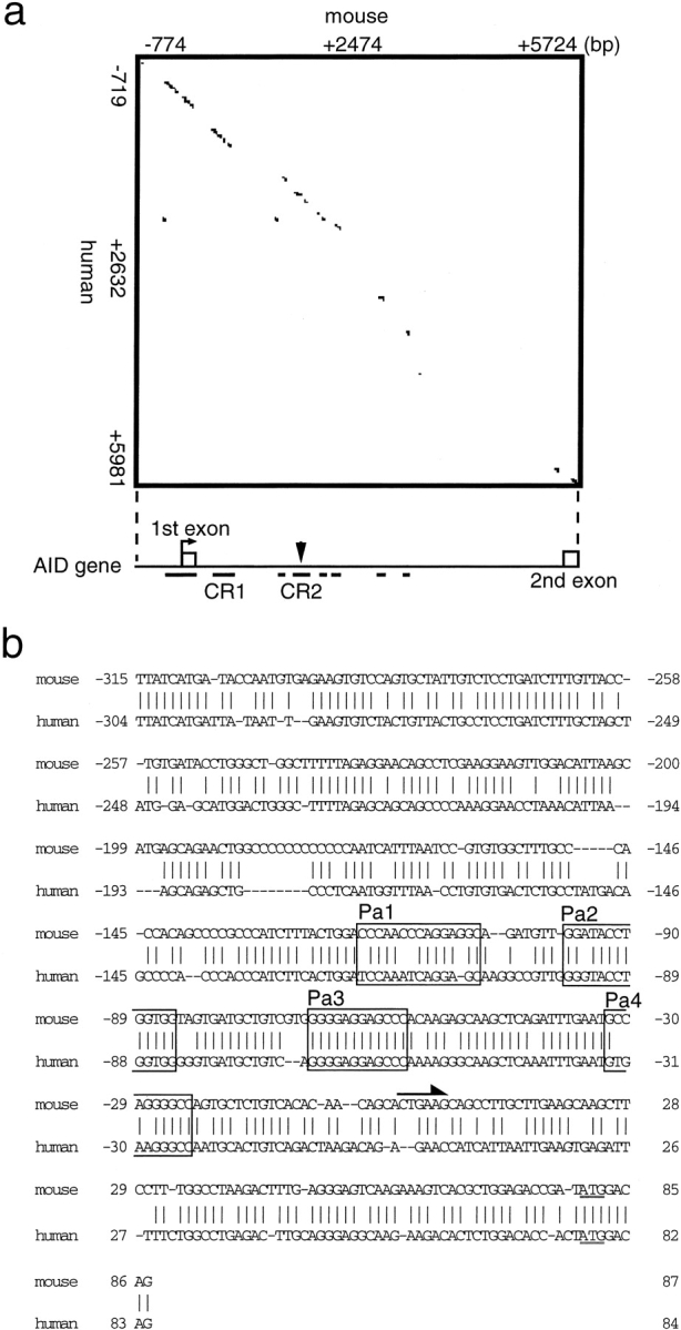
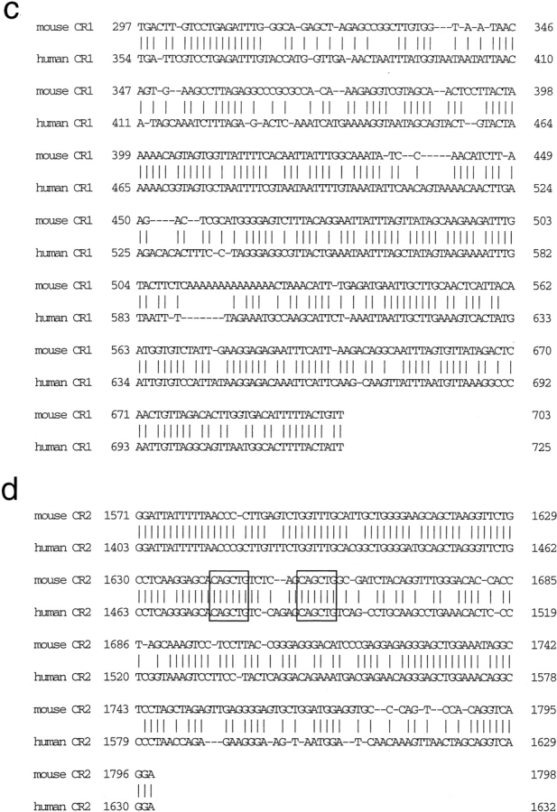
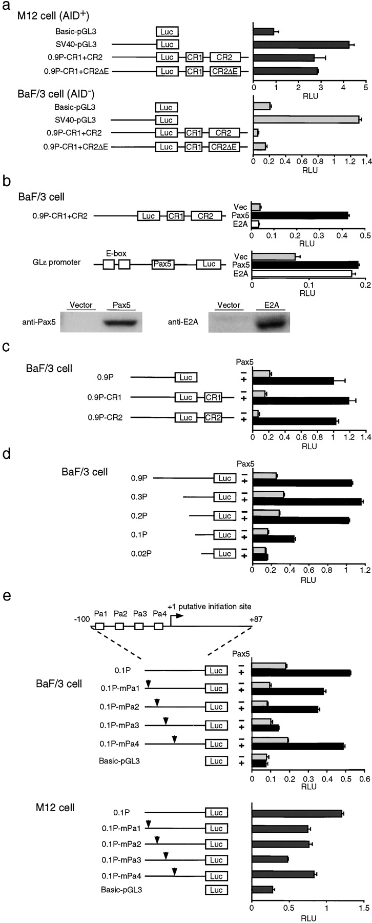
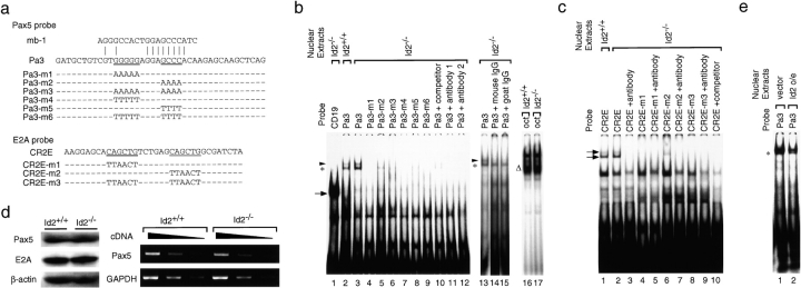
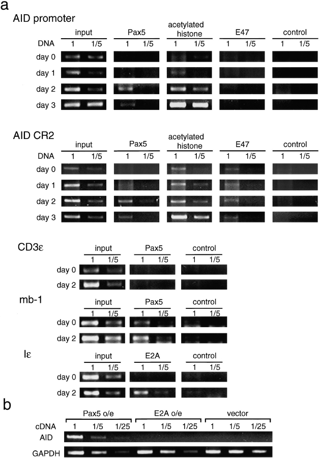
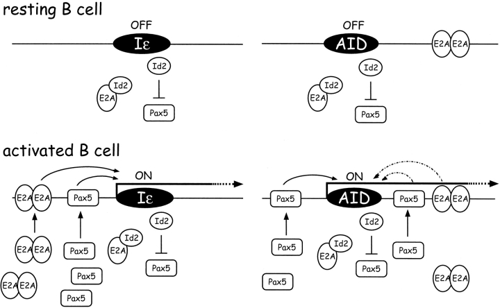
Pax5 activity is enhanced in activated B cells and is essential for class switch recombination (CSR). We show that inhibitor of differentiation (Id)2 suppresses CSR by repressing the gene expression of activation-induced cytidine deaminase (AID), which has been shown to be indispensable for CSR. Furthermore, a putative regulatory region of AID contains E2A- and Pax5-binding sites, and the latter site is indispensable for AID gene expression. Moreover, the DNA-binding activity of Pax5 is decreased in Id2-overexpressing B cells and enhanced in Id2(-/-) B cells. The kinetics of Pax5, but not E2A, occupancy to AID locus is the same as AID expression in primary B cells. Finally, enforced expression of Pax5 induces AID transcription in pro-B cell lines. Our results provide evidence that the balance between Pax5 and Id2 activities has a key role in AID gene expression.


Figures

Figure 1.
Figure 1.
Effect of enforced expression of Id2 on AID transcription. (a) A diagram of the Id2 retrovirus construct. Surface expression of truncated human CD4 was used as the marker of infected cells. (b) The expression levels of AID in splenic B cells stimulated in vitro. RNAs from Id2-transduced and control populations were subjected to RT-PCR analysis. (c) The expression levels of AID in splenic B cells stimulated in vitro. RNAs from Id2−/− and Id2+/+ B cells were subjected to RT-PCR analysis. One of three independent experiments is shown. Fivefold serial dilutions of cDNAs were amplified for the indicated transcripts. GAPDH was used as an internal control. (d) Time course analysis of the Pax5 and E2A proteins in in vitro–activated splenic B cells. Each lane contains the extract from the equal cell numbers (2 × 10⁶ cells). (e) Time course analysis of the AID expression in in vitro–activated splenic B cells. One of three independent experiments is shown. Fivefold serial dilutions of cDNAs were amplified for the indicated transcripts. GAPDH was used as an internal control.
Figure 2.
Figure 2.
Conservation of AID promoter sequence in mice and humans. (a) Dot matrix sequence comparisons of the sequences of the mouse and human AID 5′ upstream region, exon 1, intron 1, and exon 2 using a window with 33 of 50 matches. A schematic diagram of the putative regulatory regions of the AID genes in mice and humans is shown below the matrix. The open boxes indicate exons. Underlines indicate conserved sequences. (b) Alignment of 5′-flanking conserved sequences of the AID genes of mice and humans. Vertical lines indicate conserved nucleotides in the two species. The underlined sequence is the first ATG. The arrow indicates the transcription initiation site. (c) Alignment of CR1 conserved sequences of the AID genes of mice and humans. Vertical lines indicate nucleotides conserved between the two species. (d) Alignment of CR2 conserved sequences of the AID genes of mice and humans. Vertical lines indicate nucleotides conserved between the two species. The open boxes indicate the E-box sequence.
Figure 2.
Figure 2.
Conservation of AID promoter sequence in mice and humans. (a) Dot matrix sequence comparisons of the sequences of the mouse and human AID 5′ upstream region, exon 1, intron 1, and exon 2 using a window with 33 of 50 matches. A schematic diagram of the putative regulatory regions of the AID genes in mice and humans is shown below the matrix. The open boxes indicate exons. Underlines indicate conserved sequences. (b) Alignment of 5′-flanking conserved sequences of the AID genes of mice and humans. Vertical lines indicate conserved nucleotides in the two species. The underlined sequence is the first ATG. The arrow indicates the transcription initiation site. (c) Alignment of CR1 conserved sequences of the AID genes of mice and humans. Vertical lines indicate nucleotides conserved between the two species. (d) Alignment of CR2 conserved sequences of the AID genes of mice and humans. Vertical lines indicate nucleotides conserved between the two species. The open boxes indicate the E-box sequence.
Figure 3.
Figure 3.
Transcriptional activation of the putative AID promoter by Pax5. (Left) Schematic diagrams of reporter constructs containing no enhancer or promoter (Basic-pGL3), only SV40 promoter (SV40-pGL3), all the conserved sequences (0.9P-CR1+CR2), all the conserved sequences with deleted E-boxes (0.9P-CR1+CR2ΔE), promoter only (0.9P), promoter plus CR1 (0.9P-CR1), promoter plus CR2 (0.9P+CR2), various promoter deletions (0.3P, 0.2P, 0.1P, and 0.02P), various potential Pax-binding site mutations (0.1P-mPa1, 0.1P-mPa2, 0.1P-mPa3, and 0.1P-mPa4). (a, right) BaF/3 cells or M12 cells were transfected with AID–luciferase constructs (20 μg). All assays were independently performed five times in duplicate. Luciferase activity is the relative luminescence units normalized by Renilla luciferase activity. (b, right) BaF/3 cells were transfected with AID–luciferase constructs (20 μg) or Glɛ promoter constructs (20 μg) in the absence (black bars) or presence of pcDNA-Pax5 (15 μg) (black bars) or pcDNA-E47 (15 μg) (white bars). Immunoblot analysis of Pax5 and E2A proteins in 293T cells transfected with indicated expression vectors. (c) BaF/3 cells were transfected with AID–luciferase constructs (20 μg) in the absence (black bars) or presence (black bars) of pcDNA-Pax5 (15 μg). (d) BaF/3 cells were transfected with various deletion AID–luciferase constructs (20 μg) in the absence (black bars) or presence of pcDNA-Pax5 (15 μg) (black bars). (e, top column) Schematic representation of AID promoter. Boxes indicate putative Pax5-binding sequences (Pa1–Pa4). (e, left) Schematic diagrams of the mutated promoter constructs. Arrowheads indicate the mutations introduced into putative Pax5-binding sites Pa1–Pa4. (e, middle right) BaF/3 cells were transfected with AID–luciferase constructs (20 μg) in the absence (black bars) or presence (black bars) of pcDNA-Pax5 (15 μg). (e, lower right) M12 cells were transfected with the indicated AID–luciferase constructs (20 μg). Luciferase activity is the relative luminescence units normalized by Renilla luciferase activity. All assays were independently performed six times in duplicate.
Figure 4.
Figure 4.
Binding of Pax5 to the AID promoter. (a, top column, top panel) Pax5-binding sequence in the mb-1 promoter. The DNA sequences of the probes used in this assay (b) are shown. Pa3 represents the WT sequence. Underlined sequences of the WT P3 probe were changed as noted (Pa3-m1 to P3-m6). (a, bottom column) The DNA sequences of the probes used in this assay (c) are shown. CR2E represents the WT sequence. Underlined sequences of the WT CR2E probe were changed as noted (CR2E-m1 to P3-m3). (b) EMSA of nuclear extracts from splenic B cells of Id2−/− and control mice treated with 20 μg/ml LPS plus 10 ng/ml IL-4 for 3 d using the probes shown in panel a. The asterisk indicates the Pax5-containing complex. The arrowhead indicates a nonspecific band. An Oct probe was used as an internal control. One of three independent experiments is shown. (c) EMSA of nuclear extracts from splenic B cells of Id2−/− and control mice treated as described in panel b using the probes shown in panel a. The arrows indicate the E2A-containing complex. One of six independent experiments is shown. (d) The levels of Pax5 or E2A proteins and Pax5 transcripts. In vitro–stimulated B cells from Id2−/− and Id2+/+ mice were subjected to Western blotting or RT-PCR. One of three independent experiments is shown. Fivefold serial dilutions of cDNAs were amplified for the indicated transcripts. GAPDH or β-actin were used as an internal control. (d) Nuclear extracts prepared from hCD4-positive cells obtained as described in Fig. 1 were analyzed by EMSA using the P3 probe.
Figure 5.
Figure 5.
Recruitment of Pax5 to the AID promoter in activated B cells. (a) ChIP analyses at the indicated sites. Fivefold serial dilutions of input DNA and DNAs immunoprecipitated with anti-E2A, anti-Pax5, anti–acetylated histone H3, and control (antibodies) Abs were analyzed by PCR. One of three independent experiments is shown. (b) Pax5-induced endogenous AID expression in BaF/3 cells. Pax5 or E47 or empty vectors (pMX-IREShCD4) were introduced into BaF/3 cells via retroviral transfer. RNAs prepared from hCD4-positive cells were analyzed by RT-PCR. One of five independent experiments is shown.
Figure 6.
Figure 6.
Model for the regulation of AID and IgE gene expression by E2A, Pax5, and Id2.

Similar articles

Cited by

References

    1. Norton, J.D., R.W. Deed, G. Craggs, and F. Sablitzky. 1998. Id helix-loop-helix proteins in cell growth and differentiation. Trends Cell Biol. 8:58–65. - PubMed
    1. Yokota, Y., and S. Mori. 2002. Role of Id family proteins in growth control. J. Cell. Physiol. 190:21–28. - PubMed
    1. Yates, P.R., G.T. Atherton, R.W. Deed, J.D. Norton, and A.D. Sharrocks. 1999. Id helix-loop-helix proteins inhibit nucleoprotein complex formation by the TCF ETS-domain transcription factors. EMBO J. 18:968–976. - PMC - PubMed
    1. Roberts, E.C., R.W. Deed, T. Inoue, J.D. Norton, and A.D. Sharrocks. 2001. Id helix-loop-helix proteins antagonize pax transcription factor activity by inhibiting DNA binding. Mol. Cell. Biol. 21:524–533. - PMC - PubMed
    1. Zhuang, Y., P. Soriano, and H. Weintraub. 1994. The helix-loop-helix gene E2A is required for B cell formation. Cell. 79:875–884. - PubMed

Publication types