. 2003 Nov 11;100(23):13235-40.
doi: 10.1073/pnas.2135169100. Epub 2003 Oct 27.

The two rotor components of yeast mitochondrial ATP synthase are mechanically coupled by subunit delta

Affiliations

The two rotor components of yeast mitochondrial ATP synthase are mechanically coupled by subunit delta

Stéphane Duvezin-Caubet et al. Proc Natl Acad Sci U S A. .

Abstract

The mitochondrial ATP synthase is made of a membrane-integrated F0 component that forms a proton-permeable pore through the inner membrane and a globular peripheral F1 domain where ATP is synthesized. The catalytic mechanism is thought to involve the rotation of a 10-12 c subunit ring in the F0 together with the gamma subunit of F1. An important and not yet resolved question is to define precisely how the gamma subunit is connected with the c-ring. In this study, using a doxycycline-regulatable expression system, we provide direct evidence that the rest of the enzyme can assemble without the delta subunit of F1, and we show that delta-less mitochondria are uncoupled because of an F0-mediated proton leak. Based on these observations, and taking into account high-resolution structural models, we propose that subunit delta plays a key role in the mechanical coupling of the c-ring to subunit gamma.


Figures
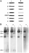
Fig. 5.
Fig. 5.
SDS/PAGE and BN/PAGE analysis of δ-less mitochondria. Mitochondria were prepared from SDC6 cells grown in a rich glucose medium (YPGA) for eight to nine generations with no addition (-) or with 10 μg/ml doxycycline plus 6 μg/ml oligomycin (+). (A) SDS/PAGE and Western blot analysis of the mitochondria with antibodies against the indicated ATP synthase subunits. (B) Mitochondrial digitonin extracts were resolved by BN/PAGE (see Materials and Methods for details of the procedure), transferred to poly(vinylidene difluoride) membranes, and analyzed by Western blot with antibodies against the indicated ATP synthase subunits.
Fig. 1.
Fig. 1.
A block in the expression of the δ subunit leads to a respiratory growth arrest. (A) Growth curves. Wild-type strain SDC22 expressing the δ subunit from its own promoter and SDC6 expressing the δ subunit from a doxycyclinerepressible promoter (tet-O) were pregrown in rich glycerol plus ethanol medium (YPEG). Fresh YPEG medium containing no or 10 μg/ml doxycycline was inoculated with these precultures (at the starting time of the growth curves shown). ⋄, SDC22 without doxycycline; ▪, SDC22 with doxycycline; •, SDC6 without doxycycline; ▾, SDC6 with doxycycline. (B) Western blot analysis. Mitochondria were prepared from the cultures shown in A at the time indicated by a dashed line (+DOX and -DOX refer to the presence or absence of doxycycline in the growth medium, respectively). The mitochondrial proteins were then analyzed by SDS/PAGE and Western blot with antibodies against the indicated ATP synthase subunits. Each lane was loaded with 10 μg of proteins.
Fig. 2.
Fig. 2.
Oxygen consumption of mitochondria. Mitochondria prepared from SDC6 grown without (A) or with (B) doxycycline (as described in the legend of Fig. 1) were assayed for oxygen consumption. The additions were 0.3 mg/ml mitochondrial proteins (M), 4 mM NADH, 400 μM ADP, 6 μg/ml oligomycin, and 3 μM CCCP.
Fig. 3.
Fig. 3.
Energization of mitochondria. Energization of the mitochondrial inner membrane was monitored by fluorescence quenching of rhodamine 123 with intact mitochondria isolated from SDC6 grown in the absence (A) or presence (B) of doxycycline (as described in the legend of Fig. 1). The additions were 0.5 μg/ml rhodamine 123, 0.3 mg/ml mitochondrial proteins (M), 10 μl of ethanol (EtOH), 6 μg/ml oligomycin, 0.2 mM potassium cyanide (KCN), and 3 μM CCCP.
Fig. 4.
Fig. 4.
Fluorescence microscopy of whole cells with a mitochondrial membrane potential probe. SDC6 was grown without (A) or with (B)10μg/ml doxycycline for 6 hr in rich galactose medium (YPGALA). The cells were then incubated for 30 min with 5 μM DASPMI and then examined by fluorescence microscopy. SDC6 cells grown without doxycycline were also incubated with 10 μM CCCP (C) to demonstrate complete dissipation of mitochondrial membrane potential. (Upper) Nomarski views. (Lower) Mitochondrial stainings. (Bar = 3 μm.)

Similar articles

Cited by

References

    1. Boyer, P. D. (1997) Annu. Rev. Biochem. 66, 717-749. - PubMed
    1. Abrahams, J. P., Leslie, A. G. W., Lutter, R. & Walker, J. E. (1994) Nature 370, 621-628. - PubMed
    1. Gibbons, C., Montgomery, M. G., Leslie, A. G. W. & Walker, J. E. (2000) Nat. Struct. Biol. 7, 1055-1061. - PubMed
    1. Stock, D., Leslie, A. G. & Walker, J. E. (1999) Science 286, 1700-1705. - PubMed
    1. Fillingame, R. H. (2000) Nat. Struct. Biol. 7, 1002-1004. - PubMed

Publication types

MeSH terms

Substances

LinkOut - more resources