

Estimation of inflammation by Doppler ultrasound: quantitative changes after intra-articular treatment in rheumatoid arthritis
- PMID: 14583566
- PMCID: PMC1754363
- DOI: 10.1136/ard.62.11.1049
Estimation of inflammation by Doppler ultrasound: quantitative changes after intra-articular treatment in rheumatoid arthritis
Abstract
Objective: To evaluate the use of ultrasound, including quantitative Doppler analysis of synovial vascularisation, before and after intra-articular treatment with glucocorticosteroids in patients with rheumatoid arthritis (RA).
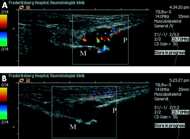
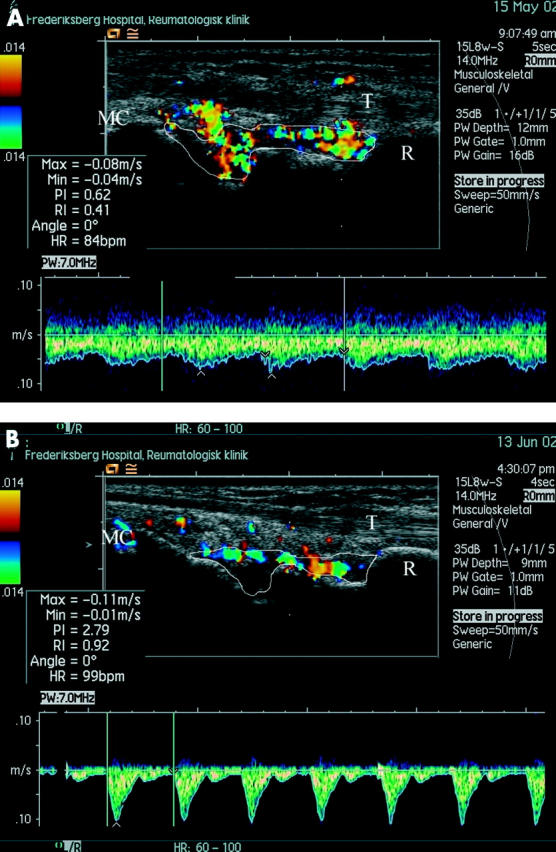
Methods: 51 patients with RA were followed prospectively after an intra-articular glucocorticosteroid injection. Disease modifying antirheumatic drug treatment was kept unchanged and no further injections given in this observation period. At baseline, disease activity was estimated clinically by target join pain on a 100 mm visual analogue scale, on which the target joint was scored 0-3 for swelling and tenderness, and by ultrasound measurements of grey scale pixels, colour Doppler pixels, and the spectral Doppler resistive index (RI) as indicators of synovial swelling and inflammation. After four weeks, the measurements were repeated on the same joint. An observer unaware of the sequence and patient number evaluated the ultrasound images.
Results: At one month follow up after the glucocorticosteroid injection, a marked decrease in the fraction of colour pixels was seen in 41/51 patients (Student's t test p<0.001). Correspondingly, the RI increased indicating a diminished flow to the synovium (Student's t test p<0.01). Both the fraction of colour pixels and the RI values corresponded with the clinical evaluation and with the subjective effect of the treatment. The synovial membrane volume estimated by total amount of pixels showed a significant decrease by 31% after treatment.
Conclusion: Ultrasound-Doppler seems to be a promising tool for the estimation of synovial activity in arthritis. After intra-articular glucocorticosteroid, changes in RI and fraction of colour pixels may both be used as quantitative measurements of the blood flow.
Figures
Similar articles
-
Spectral Doppler and resistive index. A promising tool in ultrasonographic evaluation of inflammation in rheumatoid arthritis.Acta Radiol. 2003 Nov;44(6):645-52. doi: 10.1080/02841850312331287759. Acta Radiol. 2003. PMID: 14616209
-
Contrast-enhanced power Doppler sonography of knee synovitis in rheumatoid arthritis: assessment of therapeutic response.Clin Rheumatol. 2004 Aug;23(4):285-90. doi: 10.1007/s10067-004-0878-7. Epub 2004 May 15. Clin Rheumatol. 2004. PMID: 15293087
-
Effects of treatment with etanercept (Enbrel, TNRF:Fc) on rheumatoid arthritis evaluated by Doppler ultrasonography.Ann Rheum Dis. 2003 Feb;62(2):178-81. doi: 10.1136/ard.62.2.178. Ann Rheum Dis. 2003. PMID: 12525391 Free PMC article. Clinical Trial.
-
Pro musculoskeletal ultrasonography in rheumatoid arthritis.Clin Exp Rheumatol. 2015 Jul-Aug;33(4 Suppl 92):S50-3. Epub 2015 Oct 12. Clin Exp Rheumatol. 2015. PMID: 26457337 Review.
-
Sonographic synovial vascularity of synovitis in rheumatoid arthritis.Rheumatology (Oxford). 2014 Apr;53(4):586-91. doi: 10.1093/rheumatology/ket311. Epub 2013 Sep 18. Rheumatology (Oxford). 2014. PMID: 24049097 Review.
Cited by
-
Using ultrasonography to facilitate best practice in diagnosis and management of RA.Nat Rev Rheumatol. 2009 Dec;5(12):698-706. doi: 10.1038/nrrheum.2009.227. Epub 2009 Nov 10. Nat Rev Rheumatol. 2009. PMID: 19901917 Review.
-
Power Doppler ultrasound of rheumatoid synovitis: quantification of vascular signal and analysis of interobserver variability.Skeletal Radiol. 2009 May;38(5):467-72. doi: 10.1007/s00256-009-0665-2. Epub 2009 Mar 3. Skeletal Radiol. 2009. PMID: 19255756
-
Ultrasound Color Doppler Image Segmentation and Feature Extraction in MCP and Wrist Region in Evaluation of Rheumatoid Arthritis.J Med Syst. 2016 Sep;40(9):197. doi: 10.1007/s10916-016-0552-z. Epub 2016 Jul 23. J Med Syst. 2016. PMID: 27449351
-
Power Doppler signal calibration between ultrasound machines by use of a capillary-flow phantom for pannus vascularity in rheumatoid finger joints: a basic study.Radiol Phys Technol. 2015 Jan;8(1):120-4. doi: 10.1007/s12194-014-0299-5. Epub 2014 Oct 29. Radiol Phys Technol. 2015. PMID: 25351422
-
A semiquantitative color Doppler ultrasound scoring system for evaluation of synovitis in joints of patients with blood-induced arthropathy.Insights Imaging. 2021 Sep 25;12(1):132. doi: 10.1186/s13244-021-01043-0. Insights Imaging. 2021. PMID: 34564747 Free PMC article.
