Comparative Study
. 2004 Feb;29(2):373-84.
doi: 10.1038/sj.npp.1300337.

Gender-specific gene expression in post-mortem human brain: localization to sex chromosomes

Affiliations
Comparative Study

Gender-specific gene expression in post-mortem human brain: localization to sex chromosomes

Marquis P Vawter et al. Neuropsychopharmacology. 2004 Feb.

Abstract

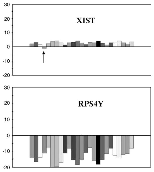
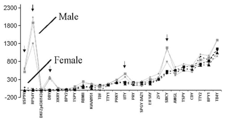
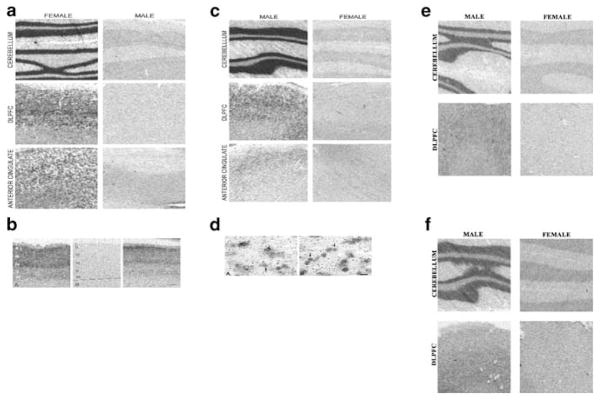
Gender differences in brain development and in the prevalence of neuropsychiatric disorders such as depression have been reported. Gender differences in human brain might be related to patterns of gene expression. Microarray technology is one useful method for investigation of gene expression in brain. We investigated gene expression, cell types, and regional expression patterns of differentially expressed sex chromosome genes in brain. We profiled gene expression in male and female dorsolateral prefrontal cortex, anterior cingulate cortex, and cerebellum using the Affymetrix oligonucleotide microarray platform. Differentially expressed genes between males and females on the Y chromosome (DBY, SMCY, UTY, RPS4Y, and USP9Y) and X chromosome (XIST) were confirmed using real-time PCR measurements. In situ hybridization confirmed the differential expression of gender-specific genes and neuronal expression of XIST, RPS4Y, SMCY, and UTY in three brain regions examined. The XIST gene, which silences gene expression on regions of the X chromosome, is expressed in a subset of neurons. Since a subset of neurons express gender-specific genes, neural subpopulations may exhibit a subtle sexual dimorphism at the level of differences in gene regulation and function. The distinctive pattern of neuronal expression of XIST, RPS4Y, SMCY, and UTY and other sex chromosome genes in neuronal subpopulations may possibly contribute to gender differences in prevalence noted for some neuropsychiatric disorders. Studies of the protein expression of these sex-chromosome-linked genes in brain tissue are required to address the functional consequences of the observed gene expression differences.


Figures

Figure 1
Figure 1
The fold change of gene expression for five males and five females samples from the DLPFC. (XIST) A predominant female expression of XIST (an exception is noted with an arrow) is shown. A higher ratio for female is shown as positive above zero, and lower expression for female is shown as below zero. (RPS4Y) The pattern for RPS4Y gene shows a predominant male expression. Both graphs depict each female sample/male combinations for five female and five male samples. The Y-axis scale ranges from −30- to 30-fold change and is calculated for each individual female/male combination in Affymetrix Microarray Suite version 4.0.1.
Figure 2
Figure 2
The mean gene expression level (y-axis) of the Y-linked probesets shown for three brain regions (large square represents anterior cingulate cortex, circle represents DLPFC, small square represents cerebellum) for male (red-solid line) and female (black-dashed line) subjects. The gene order on the x-axis is first sorted from lowest gene expression in female samples to the highest. We found consistent differences in Y expression of five genes denoted by arrows (RPS4Y, USP9Y, DBY, UTY, SMCY). We confirmed that the BPY1 gene showing above background expression in female and male brain tissue is a result of an X-homologue (VCX) expression in brain tissue. Another corresponding gene BPY2 was absent for both male and female brain tissue and confirmed to be absent by RT-PCR. Cross-hybridization of autosomal and X-linked genes with the Y probeset is a probable cause for the high expression of female brain tissue for Y-linked genes (see Table 6 for additional information).
Figure 3
Figure 3
In situ hybridization of male and female brain sections from the cerebellum, DLPFC, and anterior cingulate cortex. (a) The XIST antisense probe is shown for six sections (three males and three females) with significant pattern of labeling in neurons from female sections. (b) The XIST probe shows female DLPFC hybridization signal (left and right panels) was present in all layers of cortex with laminar-specific distribution and absent in male DLPFC layers (middle panel). In general signal was stronger in superficial layers than in deeper layers for female DLPFC. The laminar distribution and lack of hybridization in white matter suggest that XIST is prevalently expressed in neurons. (c) The 35S-XIST riboprobe silver grains reveals dense labeling of neurons and very sparse and usually absent labeling of glia. (d) The RPS4Y probe shows predominant pattern of labeling in neurons for male tissues. (e) The SMCY probe and UTY probe (f) also confirms the microarray and TaqMan/real-time PCR experiments showing a strong labeling of male cerebellum, which shows higher tissue labeling compared to both cortical regions.

References

    1. American Psychiatric Association. Diagnostic and Statistical Manual of Mental Disorders. 4. American Psychiatric Association; Washington, DC: 1994.
    1. Andreasen NC, Flaum M, Swayze V, II, O’Leary DS, Alliger R, Cohen G, et al. Intelligence and brain structure in normal individuals. Am J Psychiatry. 1993;150:130–134. - PubMed
    1. Ashburner M, Ball CA, Blake JA, Botstein D, Butler H, Cherry JM, et al. Gene ontology: tool for the unification of biology. The Gene Ontology Consortium. Nat Genet. 2000;25:25–29. - PMC - PubMed
    1. Brockdorff N, Ashworth A, Kay GF, Cooper P, Smith S, McCabe VM, et al. Conservation of position and exclusive expression of mouse Xist from the inactive X chromosome. Nature. 1991;351:329–331. - PubMed
    1. Brown CJ, Hendrich BD, Rupert JL, Lafreniere RG, Xing Y, Lawrence J, et al. The human XIST gene: analysis of a 17 kb inactive X-specific RNA that contains conserved repeats and is highly localized within the nucleus. Cell. 1992;71:527–542. - PubMed

Publication types

MeSH terms