

Characterisation of p53 status at the gene, chromosomal and protein levels in oesophageal adenocarcinoma
- PMID: 14583777
- PMCID: PMC2394414
- DOI: 10.1038/sj.bjc.6601323
Characterisation of p53 status at the gene, chromosomal and protein levels in oesophageal adenocarcinoma
Abstract
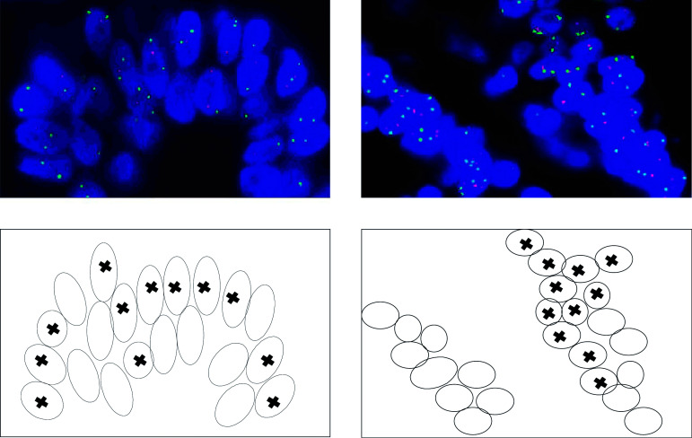
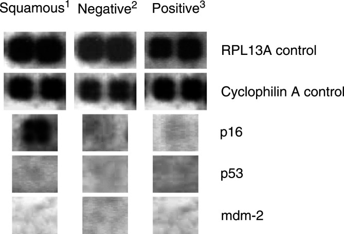
p53 mutations and loss of heterozygosity have been commonly associated with oesophageal adenocarcinoma. In this investigation, the p53 status of a Welsh population of Barrett's-associated oesophageal adenocarcinomas were fully characterised at the gene sequence, chromosomal, mRNA and protein levels. In total, 31 tumours were examined for p53 gene sequence mutations using RFLP with sequencing, allelic loss of the gene was characterised by FISH, mRNA expression by p53 pathway signalling arrays and protein levels by p53 immunohistochemistry. In all, 9.6% of adenocarcinomas harboured p53 mutations, 24% displayed p53 allelic loss and 83% exhibited p53 protein accumulation. Point mutations and deletions of the gene did not coexist within the same samples. All samples containing p53 mutations also displayed positive immunostaining; however; in the majority of cases, p53 protein accumulation developed in the absence of mutations. The gene expression analysis demonstrated no differences in p53 and mdm-2 transcription levels between the p53 immunonegative and immunopositive samples, indicating other mechanisms underlie the proteins' overexpression. In conclusion, p53 mutations and deletions do not appear to be frequent events in oesophageal adenocarcinomas; however, abnormal accumulation of the protein is present in a vast majority of cases. P53 gene mutations are not the primary cause of protein overexpression--an alternative mechanism is responsible for the positive p53 immunohistochemistry detected.
Figures
References
-
- Ashcroft M, Vousden KH (1999) Regulation of p53 stability. Oncogene 18: 7637–7643 - PubMed
-
- Battifora H (1994) p53 immunohistochemistry: a word of caution. Hum Pathol 25: 435–437 - PubMed
-
- Bian YS, Osterheld MC, Fontolliet C, Bosman FT, Benhattar J (2002) p16 inactivation by methylation of the CDKN2A promoter occurs early during neoplastic progression in Barrett's esophagus. Gastroenterology 122: 1113–1121 - PubMed
-
- Bonsing BA, Corver WE, Fleuren GJ, Cleton-Jansen AM, Devilee P, Cornelisse CJ (2000) Allelotype analysis of flow-sorted breast cancer cells demonstrates genetically related diploid and aneuploid subpopulations in primary tumors and lymph node metastases. Genes Chromosomes Cancer 282: 173–183 - PubMed
Publication types
MeSH terms
Substances
LinkOut - more resources
Full Text Sources
Medical
Research Materials
Miscellaneous
