. 2003 Nov 11;100(23):13190-5.
doi: 10.1073/pnas.1835675100. Epub 2003 Oct 29.

The crystal structures of severe acute respiratory syndrome virus main protease and its complex with an inhibitor

Affiliations

The crystal structures of severe acute respiratory syndrome virus main protease and its complex with an inhibitor

Haitao Yang et al. Proc Natl Acad Sci U S A. .

Abstract

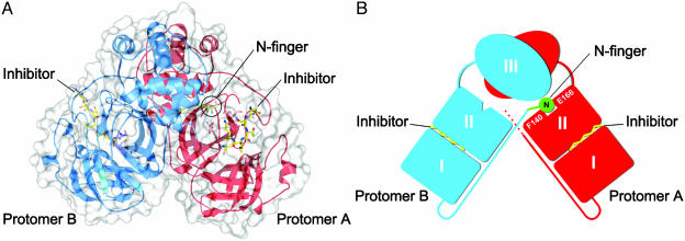
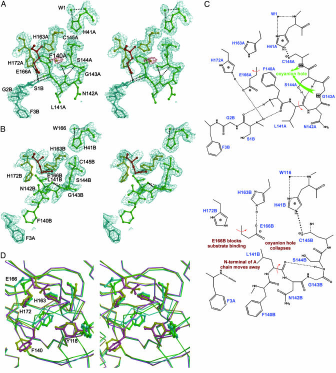
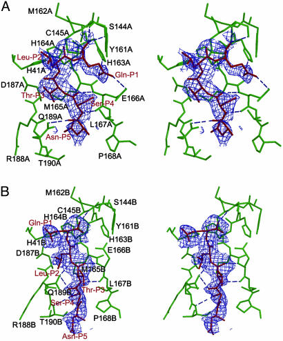
A newly identified severe acute respiratory syndrome coronavirus (SARS-CoV), is the etiological agent responsible for the outbreak of SARS. The SARS-CoV main protease, which is a 33.8-kDa protease (also called the 3C-like protease), plays a pivotal role in mediating viral replication and transcription functions through extensive proteolytic processing of two replicase polyproteins, pp1a (486 kDa) and pp1ab (790 kDa). Here, we report the crystal structures of the SARS-CoV main protease at different pH values and in complex with a specific inhibitor. The protease structure has a fold that can be described as an augmented serine-protease, but with a Cys-His at the active site. This series of crystal structures, which is the first, to our knowledge, of any protein from the SARS virus, reveal substantial pH-dependent conformational changes, and an unexpected mode of inhibitor binding, providing a structural basis for rational drug design.


Figures

Fig. 1.
Fig. 1.
The SARS-CoV Mpro dimer structure complexed with a substrate-analogue hexapeptidyl CMK inhibitor. (A) The SARS-CoV Mpro dimer structure is presented as ribbons, and inhibitor molecules are shown as ball-and-stick models. Protomer A (the catalytically competent enzyme) is red, protomer B (the inactive enzyme) is blue, and the inhibitor molecules are yellow. The N-finger residues of protomer B are green. The molecular surface of the dimer is superimposed. (B) A cartoon diagram illustrating the important role of the N-finger in both dimerization and maintenance of the active form of the enzyme.
Fig. 2.
Fig. 2.
Conformational variations in the S1 substrate-binding pocket. (A) A stereoview of the active site of protomer A built into the 1.9-Å electron density (2 Fo - Fc, contoured at 1.0 σ). The oval-shaped piece of electron density, which is red, is assigned to a water molecule. In S1 subsite of protomer A, Glu-A166 is red, His-A163 and His-A172 are yellow, and the other residues are green. Protomer B is cyan. The amino acid residues of the protein are labeled in single letters; for example, H163A stands for His-163 of monomer A (i.e., His-A163). (B) A stereo image showing the collapsed active site of protomer B built into electron density (2Fo - Fc, contoured at 1.0 σ). The oxyanion hole collapses, the N-finger of chain A is not anchored to its binding site on protomer B, Phe-B140 is directed out into bulk solvent, and Glu-B166 switches conformation to block the substrate-binding site. (C) A schematic presentation of the conformational variations and altered hydrogen-bonding networks in active sites. (Upper) The oxyanion hole (for protomer A) and N-finger of protomer B docked to its binding site. (Lower) The corresponding view of the collapsed active site in protomer B. The N-finger is not docked to its binding site, with the following consequences: (i) the oxyanion hole collapses; (ii) Phe-B140 protrudes into bulk solvent; and (iii) Glu-B166 switches conformation to block the S1 substrate-binding subsite. (D) Comparison of four SARS-CoV Mpro structures. A stereo figure is shown of the substrate-binding pocket of protomer B, with their Cα superimposed. The coloring is as follows: pH 6.0, yellow; pH 7.6, cyan; pH 8.0, green; and CMK inhibitor complex, pink. Side chains are shown as ball-and-stick models for the residues Tyr-B118, Phe-B140, Cys-B145, His-B163, Glu-B166, and His-B172. Note the dramatic conformational changes for Tyr-B118, Phe-B140, Cys-B145, and Glu-B166 when the pH changes from 6.0 to higher pH values.
Fig. 3.
Fig. 3.
The effect of pH on Mpro enzyme activity. (A) A plot of production against pH. Production is defined as the ratio of (P1 + P2):(P1 + P2 + S), where P1 and P2 represent the product of the substrate that was cleaved and S represents the substrate leftover. See Methods for further details of the pH activity assay. (B) A profile of the proteolytic reaction for determination of the enzymatic activity of SARS-CoV Mpro. Data are shown for pH 6.0. The extent of peptide cleavage was analyzed by reverse HPLC. The P1 and P2 peaks represent the product peptides that were cleaved, and the S peak represents the substrate leftover.
Fig. 4.
Fig. 4.
Molecular recognition interactions in the substrate-analogue hexapetidyl CMK inhibitor (Cbz-Val-Asn-Ser-Thr-Leu-Gln-CMK) complexed with SARS Mpro. (A) A stereoview of the substrate-binding pocket (green) in protomer A of the CMK inhibitor complex. The inhibitor molecule (red) is shown in the 2.5-Å original Fo - Fc difference electron-density map (1.5 σ). Hydrogen bonds are shown as dashed lines. The Gln-P1 is bound to the S1 substrate-specificity subsite, but Leu-P2 fails to bind at the S2 subsite (near Asp-A187), which is instead occupied by Thr-P3. The amino acid residues of the protein are labeled in single letters; for example, H163A stands for His-163 of monomer A (i.e., His-A163). (B) A stereoview of the substrate-binding pocket (green) in protomer B of the CMK inhibitor complex. The inhibitor molecule (red) is shown in the original Fo - Fc difference electron-density map (1.5 σ). The Gln-P1 does not bind to the partly collapsed S1 subsite in this protomer, but Leu-P2 and Ser-P4 are in their canonical binding sites. See text for further details.

Similar articles

Cited by

References

    1. Drosten, C., Gunther, S., Preiser, W., van der Werf, S., Brodt, H. R., Becker, S., Rabenau, H., Panning, M., Kolesnikova, L., Fouchier, R. A., et al. (2003) N. Engl. J. Med. 348, 1967-1976. - PubMed
    1. Fouchier, R. A., Kuiken, T., Schutten, M., van Amerongen, G., van Doornum, G. J., van den Hoogen, B. G., Peiris, M., Lim, W., Stohr, K. & Osterhaus, A. D. (2003) Nature 423, 240. - PMC - PubMed
    1. Ksiazek, T. G., Erdman, D., Goldsmith, C. S., Zaki, S. R., Peret, T., Emery, S., Tong, S., Urbani, C., Comer, J. A., Lim, W., et al. (2003) N. Engl. J. Med. 348, 1953-1966. - PubMed
    1. Peiris, J. S., Lai, S. T., Poon, L. L., Guan, Y., Yam, L. Y., Lim, W., Nicholls, J., Yee, W. K., Yan, W. W., Cheung, M. T., et al. (2003) Lancet 361, 1319-1325. - PMC - PubMed
    1. Marra, M. A., Jones, S. J., Astell, C. R., Holt, R. A., Brooks-Wilson, A., Butterfield, Y. S., Khattra, J., Asano, J. K., Barber, S. A., Chan, S. Y., et al. (2003) Science 300, 1399-1404. - PubMed

Publication types

MeSH terms