. 2003 Nov;23(22):7965-72.
doi: 10.1128/MCB.23.22.7965-7972.2003.

Adhesive and lateral E-cadherin dimers are mediated by the same interface

Affiliations

Adhesive and lateral E-cadherin dimers are mediated by the same interface

Regina B Troyanovsky et al. Mol Cell Biol. 2003 Nov.

Abstract

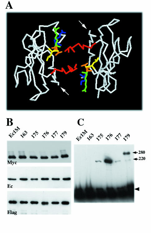
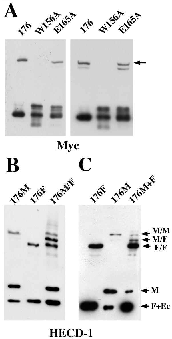
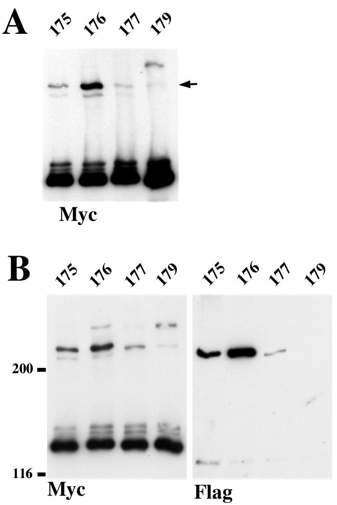
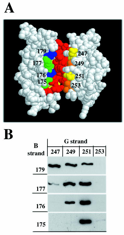
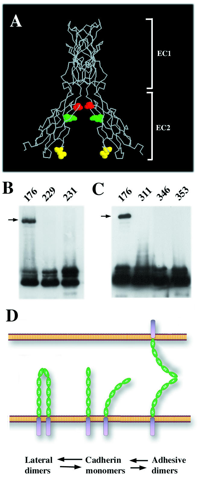
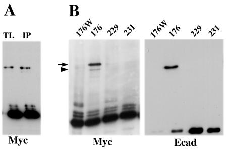
E-cadherin is a transmembrane protein that mediates Ca(2+)-dependent cell-cell adhesion. To study cadherin-cadherin interactions that may underlie the adhesive process, a recombinant E-cadherin lacking free sulfhydryl groups and its mutants with novel cysteines were expressed in epithelial A-431 cells. These cysteine mutants, designed according to various structural models of cadherin dimers, were constructed to reveal cadherin dimerization by the bifunctional sulfhydryl-specific cross-linker BM[PE0]3. Cross-linking experiments with the mutants containing a cysteine at strand B of their EC1 domains did show cadherin dimerization. By their properties these dimers correspond to those which have been characterized by co-immunoprecipitation assay. Under standard culture conditions the adhesive dimer is a dominant form. Calcium depletion dissociates adhesive dimers and promotes the formation of lateral dimers. Our data show that both dimers are mediated by the amino-terminal cadherin domain. Furthermore, the interfaces involved in both adhesive and lateral dimerization appear to be the same. The coexistence of the structurally identical adhesive and lateral dimers suggests some flexibility of the extracellular cadherin region.


Figures

FIG. 1.
FIG. 1.
Strategy of cysteine-scanning mutagenesis. (A) Backbone structure of N-cadherin EC1 domain strand dimer (25). A view is given from carboxyl termini (arrows). Strands A of the paired molecules are colored red. The side chains of residues exposed in the dimer interface corresponding to E-cadherin Leu175 (yellow), Val176 (orange), Gln177 (green), and Lys179 (blue) are shown. (B) A-431 cells stably producing Ec1M (lane Ec1M), Ec1M-C163A (lane 163), Ec1M-C163A/L175C (lane 175), Ec1M-C163A/V176C (lane 176), Ec1M-C163A/Q177C (lane 177), and Ec1M-C163A/K179C (lane 179) were cocultured with Ec1F-expressing cells and then were immunoprecipitated by an anti-myc antibody. Immunoprecipitates were analyzed for the presence of either myc-tagged mutants (Myc), coimmunoprecipitated endogenous E-cadherin (Ec) derived from both lateral and adhesive dimers, or Ec1F (Flag) derived from adhesive dimers only. (C) A-431 cells expressing Ec1M mutants as in panel B were cross-linked by BM[PEO]3, and their total lysates were analyzed by Western blotting with anti-myc. Arrows indicate two cross-linked products of 220 and 280 kDa. The arrowhead indicates the monomeric form.
FIG. 2.
FIG. 2.
A 220-kDa product represents adhesive and lateral dimers. (A) A-431 cells expressing Ec1M-C163A/V176C (lane 176) or its point mutants Ec1M-C163A/V176C/W156A (lane W156A) or Ec1M-C163A/V176C/E165A (lane E165A) were cross-linked at 2 mM (left panel) or 50 μM (right panel) calcium by BM[PEO]3 and analyzed as in Fig. 1C. In the latter case, cells were preincubated in 50 μM calcium for 10 min at 37°C to ensure that they dissociated intercellular contacts. Note that the point mutation W156A completely abolished the formation of the 220-kDa product (arrow), whereas inactivation of the presumable EC1/EC2 calcium-binding sites by E165A mutation did not significantly change the amount of the product. The second band appearing in low calcium represents a cross-linked product between myc-tagged and endogenous E-cadherin. (B) BM[PEO]3 cross-linking assay with A-431 cells expressing only Ec1M-C163A/V176C (lane 176M), only Ec1F-C163A/V176C (lane 176F), or coexpressing both these mutants (lane 176M/F). The blot was stained with HECD-1 antibody, which equally recognizes all forms of E-cadherin. (C) Ec1F-C163A/V176C- and Ec1M-C163A/V176C-expressing cells were cross-linked either separately (lanes 176F and 176M, respectively) or in coculture (lane 176M+F). The ratio between flag- and myc-tagged cells was 4:1. Arrows: M/M, cross-linked dimer in which both E-cadherin molecules are tagged by myc; M/F, myc- and flag-tagged dimer; F/F, dimer with two flag-tagged molecules; M, myc-tagged monomer; F+Ec, the band containing both flag-tagged monomer and endogenous E-cadherin. Note that the M/F product in panel C represents exclusively adhesive dimers.
FIG. 3.
FIG. 3.
(A) BM[PEO]3 cross-linking assay with different B-strand cysteine mutants (indicated as in Fig. 1C) performed at 50 μM Ca2+. Note that the relative amounts of the cross-linked 220-kDa product (arrow) are the same as under the standard calcium concentration shown in Fig. 1C. (B) Cells expressing different myc-tagged cysteine mutants (indicated above the lane as in panel A) were cocultured with the cells expressing the same mutants but tagged by flag. The ratio between flag- and myc-tagged cells was 1:1. Cocultures were immunoprecipitated with anti-myc antibody and analyzed for myc (Myc) or for flag (Flag) epitopes. Note that the relative amounts of the cross-linked adhesive dimers revealed by anti-flag staining are the same as those of cross-linked lateral dimers shown in panel A.
FIG. 4.
FIG. 4.
(A) Side view of N-cadherin EC1 domain strand dimer according to a previous study (25). The A strands of the paired molecules are red. The residues of the B (green or blue) and G (yellow or orange) strands that were subjected to mutagenesis are numbered. Note that all residues are on the same side of the dimer. In the experiment shown in panel B, cells expressing myc-tagged E-cadherin with cysteine in the B strand (vertical column) were cocultured with flag-tagged E-cadherin containing cysteine in the G strand. Cells were immunoprecipitated with anti-myc antibody, and the resulting immunoprecipitates were analyzed for myc- and flag-tagged proteins. Only anti-flag-reactive bands corresponding to cross-linked adhesive dimers are shown.
FIG. 5.
FIG. 5.
(A) Backbone structure of calcium site E-cadherin dimer (side view) according to an earlier study (21). The side chains of the EC2 domain residues Glu353 (red), Gln346 (green), and Leu311 (yellow) exposed in the dimer interface and subjected to cysteine replacement are shown. (B) A-431 cells stably producing Ec1M-C163A/V176C (lane 176), Ec1M-C163A-T229C (lane 229), and Ec1M-C163A/F231C (lane 231) were cross-linked by BM[PEO]3, and the total lysates were analyzed by Western blotting with anti-myc. (C) Ec1M mutants Ec1M-C163A/L311C (lane 311), Ec1M-C163A-Q346C (lane 346), and Ec1M-C163A/E353C (lane 353) containing cysteine mutations in the EC2 domain were analyzed as in panel B. Arrows indicate the 220-kDa cross-linked dimer. Note that cysteines in the mutants designed according to the calcium site model were unable to facilitate a cross-linking reaction. (D) The model shows formation of the adhesive and lateral dimers using the same binding site. According to this model, the extracellular cadherin region (green; EC domains are numbered) is flexible and may present a single dimerization site located at the EC1 domain in a conformation favorable either for lateral or adhesive dimerization.
FIG. 6.
FIG. 6.
Cross-linking of different cysteine Ec1M mutants in the anti-myc immunoprecipitates. (A) A-431 cells expressing the Ec1M-C163A/V176C mutant were exposed to BM[PEO]3. Their total lysate was then analyzed by Western blotting with anti-myc antibody (lane TL), or the same cells were first immunoprecipitated with anti-myc antibody; the resulting immunoprecipitate was then subjected to BM[PEO]3 cross-linking in the presence of 0.5 mM CaCl2 (lane IP). Note that the amount of the cross-linked product is approximately the same in both cases. (B) A-431 cells expressing Ec1M-C163A/V176C/W156A (lane 176W), Ec1M-C163A/V176C (lane 176), Ec1M-C163A/T229C (lane 229), and Ec1M-C163A/F231C (lane 231) were immunoprecipitated with anti-myc, and the immunoprecipitates were cross-linked by BM[PEO]3 in the absence of calcium. Samples were analyzed with myc- and E-cadherin (clone C20820)-specific antibodies. The latter recognizes endogenous cadherin coimmunoprecipitated with myc-tagged mutants. Note that the cysteine mutants unable to be cross-linked on the cell surface also cannot be cross-linked in the immunoprecipitates. Note also that, due to the Cys163 residue, endogenous E-cadherin forms a cross-linked dimer with Ec1M-C163A/V176C mutant (arrowhead) at a low calcium concentration (see also Fig. 2A).

Similar articles

Cited by

References

    1. Baumgartner, W., P. Hinterdorfer, W. Ness, A. Raab, D. Vestweber, H. Schindler, and D. Drenckhahn. 2000. Cadherin interaction probed by atomic force microscopy. Proc. Natl. Acad. Sci. USA 97:4005-4010. - PMC - PubMed
    1. Becker, J. W., H. P. Erickson, S. Hoffman, B. A. Cunningham, and G. M. Edelman. 1989. Topology of cell adhesion molecules. Proc. Natl. Acad. Sci. USA 86:1088-1092. - PMC - PubMed
    1. Bibert, S., M. Jaquinod, E. Concord, C. Ebel, E. Hewat, C. Vanbelle, P. Legrand, M. Weidenhaupt, T. Vernet, and D. Gulino-Debrac. 2002. Synergy between extracellular modules of vascular endothelial cadherin promotes homotypic hexameric interactions. J. Biol. Chem. 277:12790-12801. - PubMed
    1. Blaschuk, O. W., and T. M. Rowlands. 2002. Plasma membrane components of adherens junctions. Mol. Membr. Biol. 19:75-80. - PubMed
    1. Boggon, T. J., J. Murray, S. Chappuis-Flament, E. Wong, B. M. Gumbiner, and L. Shapiro. 2002. C-cadherin ectodomain structure and implications for cell adhesion mechanisms. Science 296:1308-1313. - PubMed

Publication types

LinkOut - more resources