. 2003 Nov;23(22):7992-8007.
doi: 10.1128/MCB.23.22.7992-8007.2003.

Regulation of the human MSH6 gene by the Sp1 transcription factor and alteration of promoter activity and expression by polymorphisms

Affiliations

Regulation of the human MSH6 gene by the Sp1 transcription factor and alteration of promoter activity and expression by polymorphisms

Isabella Gazzoli et al. Mol Cell Biol. 2003 Nov.

Abstract

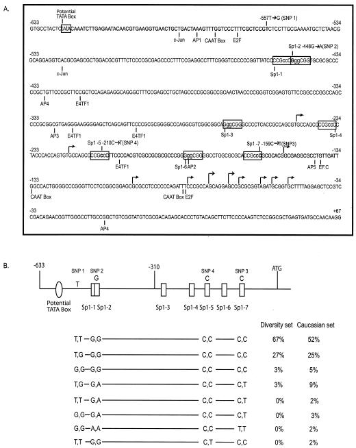
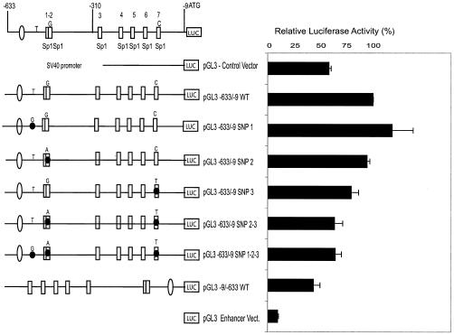
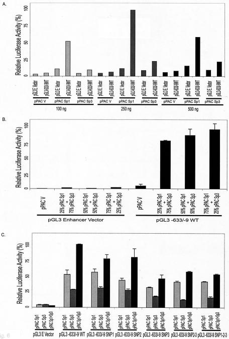
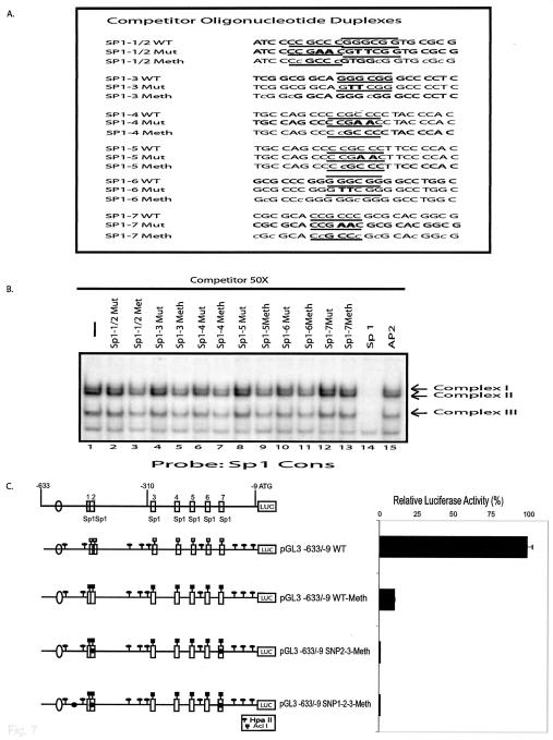
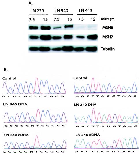
Defects in human DNA mismatch repair have been reported to underlie a variety of hereditary and sporadic cancer cases. We characterized the structure of the MSH6 promoter region to examine the mechanisms of transcriptional regulation of the MSH6 gene. The 5'-flanking region of the MSH6 gene was found to contain seven functional Sp1 transcription factor binding sites that each bind Sp1 and Sp3 and contribute to promoter activity. Transcription did not appear to require a TATA box and resulted in multiple start sites, including two major start sites and at least nine minor start sites. Three common polymorphisms were identified in the promoter region (-557 T-->G, -448 G-->A, and -159 C-->T): the latter two were always associated, and each of these functionally inactivated a different Sp1 site. The polymorphic allele -448 A -159 T was demonstrated to be a common Caucasian polymorphism found in 16% of Caucasians and resulted in a five-Sp1-site promoter that had 50% less promoter activity and was more sensitive to inactivation by DNA methylation than the more common seven Sp1 site promoter allele, which was only partially inactivated by DNA methylation. In cell lines, this five-Sp1-site polymorphism resulted in reduced MSH6 expression at both the mRNA and protein level. An additional 2% of Caucasians contained another polymorphism, -210 C-->T, which inactivated a single Sp1 site that also contributes to promoter activity.


Figures

FIG. 1.
FIG. 1.
Identification of cis-acting elements and SNPs in the MSH6 promoter. (A) The sequence of nucleotides −633 to +167 relative to the ATG of the MSH6 gene is shown. The Sp1 sites and the potential TATA box analyzed in the present study are indicated in boxes. Other potential transcription factor binding sites are indicated below the sequence. The major and minor transcription start sites are indicated by the tall and short arrows, respectively. The nucleotides altered by the 2-bp substitution mutations used in the analysis of the Sp1 sites are indicated in lowercase, and the four SNPs identified are indicated above the sequence. (B) Schematic representation of the MSH6 promoter region, with the most prevalent nucleotide at each polymorphic site indicated above the promoter diagram. Below the promoter diagram are indicated the eight different genotypes of the MSH6 promoter region identified by analyzing the SNPs present at, from left to right, nucleotides −557 (SNP 1), −448 (SNP 2), −210 (SNP 4), and −159 (SNP 3), respectively. Also indicated are the percentages of the samples from the human diversity set of DNAs (first 100 DNAs) and the Caucasian DNAs found to have each genotype.
FIG. 2.
FIG. 2.
Transcription of the MSH6 gene initiates from multiple start sites. Genescan analysis of 5′-RACE-PCR products was performed as described in Materials and Methods. Below the chromatogram is indicated a molecular size scale in base pairs determined by using appropriate markers (TAMRA).
FIG. 3.
FIG. 3.
EMSA of the seven Sp1 sites found in the MSH6 promoter region. (A) Sequences of the competitor and probe DNAs used in gel mobility shift assays. Only the sequence of the top strand of the oligonucleotide duplexes is indicated and, where relevant, the MSH6 sequence coordinates are also indicated. The Sp1 binding consensus sequences (GGGCGG) and inverted complement sequences (CCGCCC) are indicated by the overlining and underlining, respectively. The numbering system for each of the Sp1 sites is as defined in Fig. 1B. Each Sp1 sequence is wild type except that Sp1-1WT/2SNP 2 refers to the sequence containing the −448A SNP 2, Sp1-1Mut/2SNP 2 refers to the sequence containing the 2-bp substitution mutation in the Sp1-1 site and the −448A SNP 2, Sp1-1WT/2Mut refers to the sequence containing the 2-bp substitution mutation in the Sp1-2 site, and Sp1-7SNP 3 refers to the sequence containing the −159T SNP 3. See Fig. 1A for the sequences that are changed by these nucleotide substitutions. (B) Gel mobility shift assays performed with HeLa cell extract, radioactively labeled Sp1 consensus sequence as a probe, and a 50-fold molar excess of the indicated competitor DNAs. The arrows indicate the three specific protein-DNA complexes formed. (C) Effect of anti-Sp1 and anti-Sp3 antibodies on gel shift assays. Assays were performed with HeLa cell extract, and the radioactive substrate is indicated above each set of three lanes. After complex formation, the antibody indicated above each individual lane was added. The arrows indicate the three specific protein-DNA complexes formed and the position of the supershifted (S.S.) species formed after the addition of antibodies.
FIG. 4.
FIG. 4.
Effects of deletion and two-base substitution mutations on MSH6 promoter activity in transient-transfection assays. The wild-type MSH6 promoter and different mutant derivatives were inserted into the pGL3 enhancer luciferase reporter vector, and the promoter activity of each construct was assayed after transfection into either HeLa or CHO cells. (A) Analysis of deletion mutant derivatives by transfection into HeLa cells. (B) Analysis of individual 2-bp substitution mutations after transfection into HeLa cells. (C) Analysis of combinations of different 2-bp substitution mutations after transfection into CHO cells. The boxes and circles indicate the Sp1 sites and a potential TATA box as indicated in Fig. 1, respectively, with the open boxes and circles indicating the wild-type sequences and the solid boxes and circles indicating the presence of 2-bp substitution mutations: M TATA is a 2-bp substitution in the TATA box, M 1-2 indicates 2-bp substitutions in both the Sp1-1 and Sp1-2 sites, and M 3 through M 7 indicate 2-bp substitutions in the Sp1-3 through Sp1-7 sites, respectively (see Materials and Methods and Fig. 1A for details). The DNA present in the deletion mutants is indicated by the horizontal line and structural features (boxes and circles) present in the diagram: pGL3 WT contains MSH6 nucleotides −633 to −9, pGL3 Δ1 contains MSH6 nucleotides −490 to −9, pGL3 Δ2 contains MSH6 nucleotides −318 to −9, pGL3 Δ3 contains MSH6 nucleotides −248 to −9, pGL3 Δ4 contains MSH6 nucleotides −221 to −9, pGL3 Δ5 contains MSH6 nucleotides −120 to −9, pGL3 Δ6 contains MSH6 nucleotides −633 to −252, pGL3 Δ7 contains MSH6 nucleotides −633 to −193, and pGL3 Δ8 does not contain any MSH6 sequences.
FIG. 5.
FIG. 5.
Effect of SNPs on the activity of the MSH6 promoter. The pGL3 wild-type MSH6 promoter reporter vector containing MSH6 nucleotides −633 to −9 and derivatives containing single base substitutions corresponding to the −557G (SNP 1), −448A (SNP 2), and −159T (SNP 3) SNPs or combinations of these substitutions were analyzed for promoter activity after transfection into CHO cells. The presence of base substitutions of interest is indicated by the nucleotides indicated above each promoter diagram and by the black circle on the relevant structural feature of the promoter diagram when an SNP is present. The pGL3 control vector contains the simian immunodeficiency virus 40 promoter and the vector pGL3-9/-633WT contains the MSH6 promoter region in reverse orientation.
FIG. 6.
FIG. 6.
Activation of the MSH6 promoter by Sp1 and Sp3 proteins in SL2 cells. The effect of Sp1 and Sp3 transcription factors on the expression of luciferase driven by the MSH6 promoter was analyzed by cotransfection of the pGL3 vector or the pGL3-633/-9 vector containing the wild-type MSH6 promoter and either Sp1 or Sp3 expression plasmids into Drosophila SL2 cells which lack endogenous Sp1 and Sp3. (A) Drosophila SL2 cells were cotransfected with the indicated amounts of the pPAC-Sp1, pPAC-Sp3, or pPAC (empty) expression vectors and 0.4 μg of either the promoterless reporter construct pGL3-Basic vector or the pGL3-633/-9 vector containing the wild-type MSH6 promoter. (B) Drosophila SL2 cells were cotransfected with 0.4 μg of either the promoterless pGL3-Enhancer vector or the pGL3-633/-9 MSH6 promoter vector and 0.25 μg of either the pPAC empty vector or the indicated mixture of the pPAC-Sp1 and pPAC-Sp3 expression vectors. (C) Drosophila SL2 cells were cotransfected with 0.4 μg of either the promoterless pGL3-Enhancer vector, the pGL3-633/-9 MSH6 promoter vector, or derivatives of pGL3-633/-9 containing the indicated SNPs (see Fig. 5 for details about the SNP containing plasmids) and 0.25 μg of either the pPAC-Sp1, pPAC-Sp3, or an equimolar mixture of the pPAC-Sp1 and pPAC-Sp3 expression vectors.
FIG. 7.
FIG. 7.
Effect of DNA methylation on the MSH6 promoter Sp1 sites and promoter activity. (A) Sequences of the competitor DNAs used in gel mobility shift assays. Only the sequence of the top strand of the oligonucleotide duplexes is indicated (see Fig. 3 for more details). The Sp1 binding consensus sequences (GGGCGG) and inverted complement sequences (CCGCCC) are indicated by the overlining and underlining, respectively. The numbering system for each of the Sp1 sites is as defined in Fig. 1B. The wild-type sequences are indicated (WT). The mutant sequence Sp1-1/2Mut refers to the sequence containing the indicated 2-bp substitution mutations in both the Sp1-1 and Sp1-2 sites, whereas otherwise “Mut” refers to the presence of the indicated 2-bp substitution mutations in Sp1 sites Sp1-3 through Sp1-7. See Fig. 1A for sequences that are changed by these nucleotide substitutions. “Meth” refers to the wild-type sequences that have been methylated in vitro at the Cs designated in lowercase. (B) Gel mobility shift assays performed with HeLa cell extract, radioactively labeled Sp1 consensus sequence as probe (see Fig. 3A), and a 50-fold molar excess of the indicated competitor DNAs. The arrows indicate the three specific protein-DNA complexes formed. (C) The pGL3 wild-type MSH6 promoter reporter vector containing MSH6 nucleotides −633 to −9 and methylated (Meth) versions of this plasmid or derivatives containing single base substitutions corresponding to the −448A (SNP 2) and −159T (SNP 3) SNPs or the −557G (SNP 1), −448A (SNP 2), and −159T (SNP 3) SNPs were analyzed for promoter activity after transfection into HeLa cells. The positions of the HpaII and AciI sites that are substrates for DNA methylation are indicated by the symbols above the line diagram of each promoter, and the presence and position of the SNPs are indicated by the black circles on each line diagram.
FIG. 8.
FIG. 8.
Analysis of MSH6 expression in glioblastoma cell lines. (A) Western blot analysis of MSH6, MSH2, and tubulin expression. Cell extracts were prepared from the LN229, LN340, and LN443 and analyzed by Western blotting as described in Materials and Methods. Portions (7.5 and 15 μg) of each extract were analyzed as indicated above the individual lanes. The MSH6, MSH2, and tubulin bands are indicated on the right side of the gel. (B) Sequence analysis of MSH6 mRNA expression from the five and seven Sp1 site promoters. Genomic DNA and cDNA from LN340 cells were prepared and sequenced as described in Materials and Methods. LN340 cells are heterozygous for two different MSH6 alleles: one allele is the seven Sp1 site promoter allele −557G −448G −159C 186A (exon 1) 642C (exon 4), and the other allele is the five Sp1 site promoter allele −557G −448A −159T 186C (exon 1) 642T (exon 4). The sequences on the left cover the region around nucleotide 186 from a control (186C) DNA and LN340 genomic and cDNA as indicated. The sequences on the right cover the region around nucleotide 642 from a control (642C) DNA and LN340 genomic and cDNA as indicated. Reduction of the relative levels of the 186C and 642T nucleotides linked to the five Sp1 site promoter compared to the levels of the 186A and 642C nucleotides linked to the seven Sp1 site promoter is seen in the cDNA relative to the genomic DNA.

References

    1. Acharya, S., T. Wilson, S. Gradia, M. F. Kane, S. Guerrette, G. T. Marsischky, R. Kolodner, and R. Fishel. 1996. hMSH2 forms specific mispair-binding complexes with hMSH3 and hMSH6. Proc. Natl. Acad. Sci. USA 93:13629-13634. - PMC - PubMed
    1. Akiyama, Y., H. Nagasaki, T. Nakajima, H. Sakai, T. Nomizu, and Y. Yuasa. 2001. Infrequent frameshift mutations in the simple repeat sequences of hMLH3 in hereditary nonpolyposis colorectal cancers. Jpn. J. Clin. Oncol. 31:61-64. - PubMed
    1. Andrew, S. D., P. J. Delhanty, L. M. Mulligan, and B. G. Robinson. 2000. Sp1 and Sp3 transactivate the RET proto-oncogene promoter. Gene 256:283-291. - PubMed
    1. Aslam, F., L. Palumbo, L. H. Augenlicht, and A. Velcich. 2001. The Sp family of transcription factors in the regulation of the human and mouse MUC2 gene promoters. Cancer Res. 61:570-576. - PubMed
    1. Bearzatto, A., M. Szadkowski, P. Macpherson, J. Jiricny, and P. Karran. 2000. Epigenetic regulation of the MGMT and hMSH6 DNA repair genes in cells resistant to methylating agents. Cancer Res. 60:3262-3270. - PubMed

Publication types

MeSH terms