. 2003 Nov;23(22):8019-29.
doi: 10.1128/MCB.23.22.8019-8029.2003.

Pyk2- and Src-dependent tyrosine phosphorylation of PDK1 regulates focal adhesions

Affiliations

Pyk2- and Src-dependent tyrosine phosphorylation of PDK1 regulates focal adhesions

Yoshihiro Taniyama et al. Mol Cell Biol. 2003 Nov.

Abstract

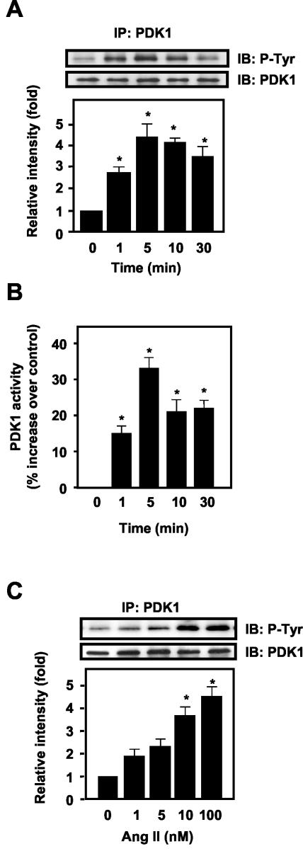
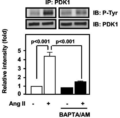
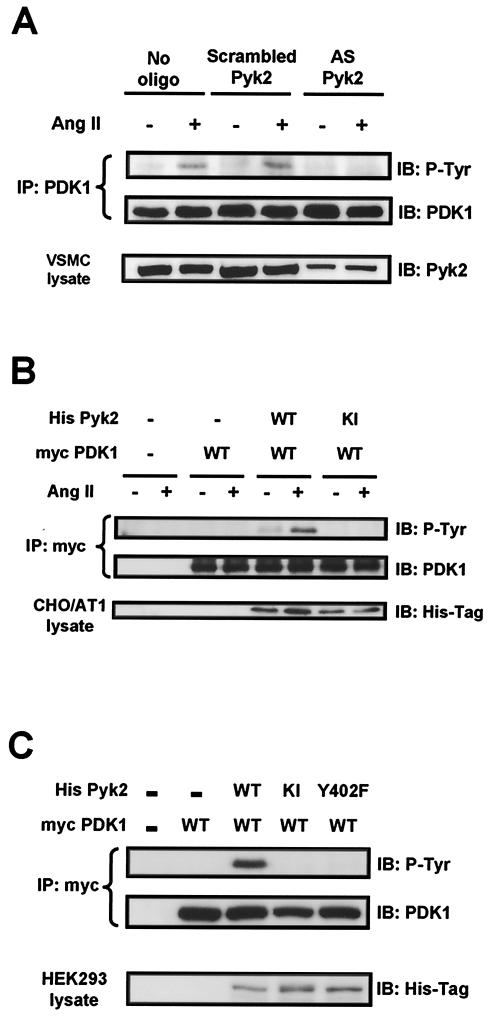
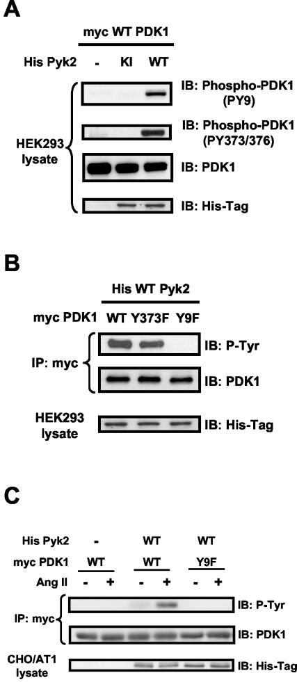
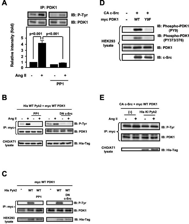
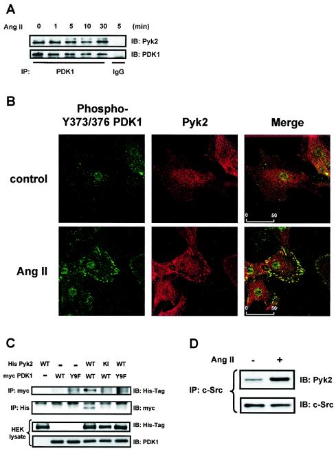
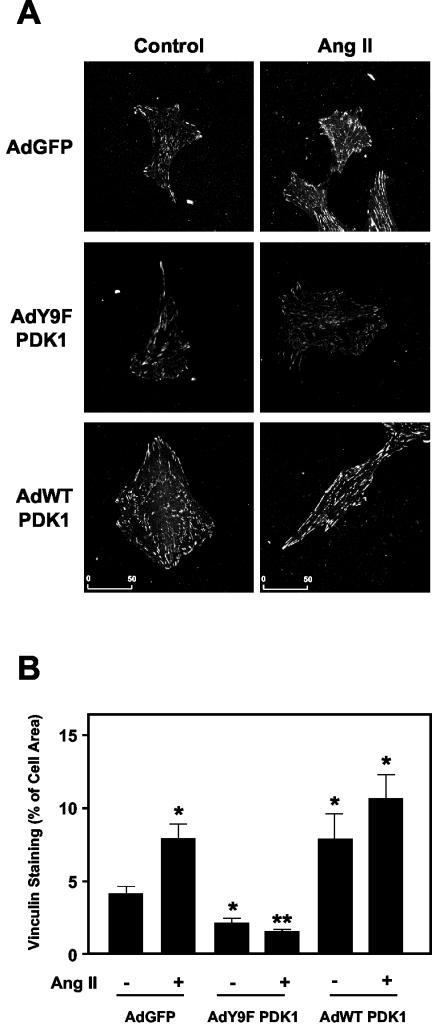
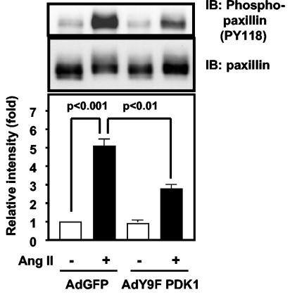
3-Phosphoinositide-dependent protein kinase 1 (PDK1) is a signal integrator that activates the AGC superfamily of serine/threonine kinases. PDK1 is phosphorylated on tyrosine by oxidants, although its regulation by agonists that stimulate G-protein-coupled receptor signaling pathways and the physiological consequences of tyrosine phosphorylation in this setting have not been fully identified. We found that angiotensin II stimulates the tyrosine phosphorylation of PDK1 in vascular smooth muscle in a calcium- and c-Src-dependent manner. The calcium-activated tyrosine kinase Pyk2 acts as a scaffold for Src-dependent phosphorylation of PDK1 on Tyr9, which permits phosphorylation of Tyr373 and -376 by Src. This critical function of Pyk2 is further supported by the observation that Pyk2 and tyrosine-phosphorylated PDK1 colocalize in focal adhesions after angiotensin II stimulation. Importantly, infection of smooth muscle cells with a Tyr9 mutant of PDK1 inhibits angiotensin II-induced tyrosine phosphorylation of paxillin and focal adhesion formation. These observations identify a novel interaction between PDK1 and Pyk2 that regulates the integrity of focal adhesions, which are major compartments for integrating signals for cell growth, apoptosis, and migration.


Figures

FIG.1.
FIG.1.
Effect of Ang II on tyrosine phosphorylation of PDK1 in VSMCs. VSMCs were stimulated with Ang II for the indicated times and concentrations and prepared for immunoprecipitation analysis or PDK1 activity assay as described in Materials and Methods. (A) Cells were stimulated with 100 nM Ang II for the indicated times, immunoprecipitated (IP) with anti-PDK1 antibody, and analyzed by Western blot analysis (IB). (B) Immunoprecipitates prepared as for panel A were assayed for activity against the PDK1 substrate PDKtide. (C) VSMCs were stimulated with various concentrations of Ang II (1 to 100 nM) for 5 min. In panels A and C, the top panels are representative immunoblots of Ang II-induced tyrosine phosphorylation of PDK1. The bottom panels represent averaged data quantified by densitometry of immunoblots, expressed as fold increases in phosphorylation. In all panels, values are means ± SE for four independent experiments. *, P < 0.05 versus control.
FIG. 2.
FIG. 2.
Role of intracellular calcium in Ang II-induced tyrosine phosphorylation in VSMCs. VSMCs were preincubated with BAPTA/AM (20 μM) for 30 min before exposure to Ang II (100 nM for 5 min). Top panels, representative immunoblots; bottom panels, averaged data quantified by densitometry of immunoblots, expressed as fold increases in phosphorylation. Values are means ± SE for three independent experiments.
FIG. 3.
FIG. 3.
Role of Pyk2 in PDK1 tyrosine phosphorylation. (A) VSMCs were transfected with either no oligonucleotide (no oligo), scrambled oligonucleotide for Pyk2 (scrambled Pyk2), or antisense oligonucleotide for Pyk2 (AS Pyk2) and were stimulated with Ang II (100 nM for 5 min). Lysates were immunoprecipitated (IP) with anti-PDK1 antibody, followed by immunoblotting (IB) with antiphosphotyrosine antibody (P-Tyr) (top) or anti-PDK1 antibody (middle). Blots are representative of three independent experiments. (B) CHO/AT1 cells were cotransfected with the indicated expression vectors and treated with either 100 nM Ang II (+) or vehicle (−) for 5 min. Lysates were immunoprecipitated with anti-Myc antibody, followed by immunoblotting with antiphosphotyrosine antibody (P-Tyr) (top) or anit-PDK1 antibody (middle). Immunoblotting of lysates with anti-His antibody shows the expression of His-tagged Pyk2 proteins (bottom). (C) HEK293 cells were cotransfected with the indicated expression vectors, and lysates were subjected to immunoprecipitation with anti-Myc antibody, followed by immunoblotting with antiphosphotyrosine antibody (P-Tyr) (top) or anti-PDK1 antibody (middle). Immunoblotting of lysates with anti-His antibody shows the expression of His-tagged Pyk2 proteins (bottom). For all panels, immunoblots are representative of three independent experiments.
FIG.4.
FIG.4.
Pyk2-dependent phosphorylation sites on PDK1. (A) HEK293 cells were cotransfected with the indicated expression vectors, and lysates were subjected to Western blot analysis (IB) with phosphorylation-site-specific anti-PDK1 antibodies (PY9 and PY373/376), total PDK-1 antibody, or anti-His antibody. (B) HEK293 cells were cotransfected with the indicated expression vectors, and lysates were subjected to immunoprecipitation (IP) with anti-Myc antibody, followed by immunoblotting with antiphosphotyrosine antibody (P-Tyr) or anti-PDK1 antibody. Separate aliquots of the lysates were immunoblotted with anti-His antibody to confirm equal expression of Pyk2. (C) CHO/AT1 cells were cotransfected with the indicated expression vectors and treated with either vehicle (−) or Ang II (+) (100 nM for 5 min). Lysates were immunoprecipitated with anti-Myc antibody, followed by immunoblotting with antiphosphotyrosine antibody (P-Tyr) (top) oranti-PDK1 antibody (middle). Immunoblotting of lysates with anti-His antibody demonstrates the expression of His-tagged Pyk2 proteins (bottom). For all panels, immunoblots are representative of three independent experiments.
FIG.5.
FIG.5.
Role of c-Src in Pyk2-dependent tyrosine phosphorylation of PDK1. (A) VSMCs were preincubated with PP1 (20 μM) for 30 min before exposure to Ang II (100 nM for 5 min). The lysates were immunoprecipitated (IP) with anti-PDK1 antibody, followed by immunoblotting (IB) with antiphosphotyrosine antibody (P-Tyr) or anti-PDK1 antibody. Top, representative immunoblots; bottom, averaged data quantified by densitometry of immunoblots, expressed as fold increases in phosphorylation. Values are means ± SE for three independent experiments. (B) CHO/AT1 cells were cotransfected with the indicated expression vectors and treated with either vehicle (−) or Ang II (+) (100 nM for 5 min). Left panels, some cells were pretreated with PP1 (10 μM for 30 min); right panels, some cells were transfected with dominant-negative (DN) c-Src together with WT His-Pyk2 and WT Myc-PDK1. Lysates were immunoprecipitated with anti-Myc antibody, followed by immunoblotting with antiphosphotyrosine antibody (P-Tyr) (top) or anti-PDK1 antibody (middle). Immunoblotting of lysates with anti-His antibody demonstrates the expression of His-tagged Pyk2 proteins (bottom). (C) HEK293 cells were cotransfected with the indicated expression vectors. Left panels, some cells were pretreated with PP1 (10 μM for 30 min); right panels, some cells were transfected with dominant-negative c-Src together with WT His-Pyk2 and WT Myc-PDK1. Cells were lysed and subjected to immunoprecipitation and immunoblotting. Immunoblotting of lysates with anti-His antibody demonstrates the expression of His-tagged Pyk2 proteins (bottom). (D) HEK293 cells were cotransfected with the indicated expression vectors and lysed, and the lysates were subjected to Western blot analysis. (E) CHO/AT1 cells were cotransfected with the indicated expression vectors and treated with either vehicle (−) or Ang II (+) (100 nM for 5 min). Lysates were immunoprecipitated with anti-Myc antibody, followed by immunoblotting with the indicated antibodies. All panels show representative immunoblots of three independent experiments.
FIG. 6.
FIG. 6.
Pyk2 association with PDK1. (A) VSMCs were stimulated with 100 nM Ang II for the indicated times, and lysates were immunoprecipitated (IP) with anti-PDK1 antibody, followed by immunoblotting (IB) with Pyk2 antibody. (B) VSMCs grown on coverslips were treated with either vehicle (control) or Ang II (100 nM for 5 min) and fluorescently stained for PDK1 phosphorylated on Y373-Y376 (green) (left panels) or Pyk2 (red) (middle panels). Right panels, merged images showing colocalization (yellow). Scale bar = 50 μm. (C) HEK293 cells were cotransfected with the indicated expression vectors, and lysates were subjected to immunoprecipitation and immunoblotting. Separate aliquots of the lysates were immunoblotted with anti-His antibody or anti-PDK1 antibody to confirm equal expression of Pyk2 or PDK1. (D) VSMCs were stimulated with Ang II (100 nM for 5 min), and lysates were immunoprecipitated with anti-c-Src antibody, followed by immunoblotting with Pyk2 antibody (top). Equal amounts of c-Src protein in the immunoprecipitates were confirmed by immunoblotting with anti-c-Src antibody (bottom). For panels A, C, and D, the data are representative immunoblots of three independent experiments.
FIG. 7.
FIG. 7.
Role of PDK1 tyrosine phosphorylation in Ang II-induced FA formation in VSMCs. (A) VSMCs were infected with either control adenovirus expressing GFP (AdGFP), virus expressing WT PDK1 (AdWT PDK1), or virus expressing Y9F PDK1 (AdY9F PDK1). Cells were treated with either vehicle (control) or Ang II (100 nM for 5 min). The results are representative of two independent experiments in which 30 cells in 10 fields were observed. Scale bar = 50 μm. (B) Quantification of vinculin staining in the cells described for panel A. A total of 7 to 10 cells from each group were analyzed as described in Materials and Methods, and the area of vinculin staining was expressed as a percentage of the total cell area. *, significant difference from GFP alone (P < 0.05); **, significant difference from Ang II alone in GFP-infected cells (P < 0.01).
FIG. 8.
FIG. 8.
Role of PDK1 tyrosine phosphorylation in Ang II-induced paxillin phosphorylation. VSMCs were infected with the indicated adenoviruses, treated with Ang II (100 nM for 5 min), lysed, and immunoblotted (IB) with phospho (pY118)-paxillin antibody. Top panels, representative immunoblots; bottom panel, averaged data quantified by densitometry, expressed as fold increases in phosphorylation. Values are means ± SE for four independent experiments.

Similar articles

Cited by

References

    1. Alessi, D. R., M. Deak, A. Casamayor, F. B. Caudwell, N. Morrice, D. G. Norman, P. Gaffney, C. B. Reese, C. N. MacDougall, D. Harbison, A. Ashworth, and M. Bownes. 1997. 3-Phosphoinositide-dependent protein kinase-1 (PDK1): structural and functional homology with the Drosophila DSTPK61 kinase. Curr. Biol. 7:776-789. - PubMed
    1. Alessi, D. R., S. R. James, C. P. Downes, A. B. Holmes, P. R. Gaffney, C. B. Reese, and P. Cohen. 1997. Characterization of a 3-phosphoinositide-dependent protein kinase which phosphorylates and activates protein kinase B alpha. Curr. Biol. 7:261-269. - PubMed
    1. Anderson, K. E., J. Coadwell, L. R. Stephens, and P. T. Hawkins. 1998. Translocation of PDK-1 to the plasma membrane is important in allowing PDK-1 to activate protein kinase B. Curr. Biol. 8:684-691. - PubMed
    1. Avraham, H., S. Y. Park, K. Schinkmann, and S. Avraham. 2000. RAFTK/Pyk2-mediated cellular signalling. Cell Signal. 12:123-133. - PubMed
    1. Berk, B. C. 1999. Angiotensin II signal transduction in vascular smooth muscle: pathways activated by specific tyrosine kinases. J. Am. Soc. Nephrol. 10(Suppl. 11):S62-S68. - PubMed

Publication types

MeSH terms