. 2003 Nov;23(22):8172-88.
doi: 10.1128/MCB.23.22.8172-8188.2003.

Retinoblastoma tumor suppressor: analyses of dynamic behavior in living cells reveal multiple modes of regulation

Affiliations

Retinoblastoma tumor suppressor: analyses of dynamic behavior in living cells reveal multiple modes of regulation

Steven P Angus et al. Mol Cell Biol. 2003 Nov.

Abstract

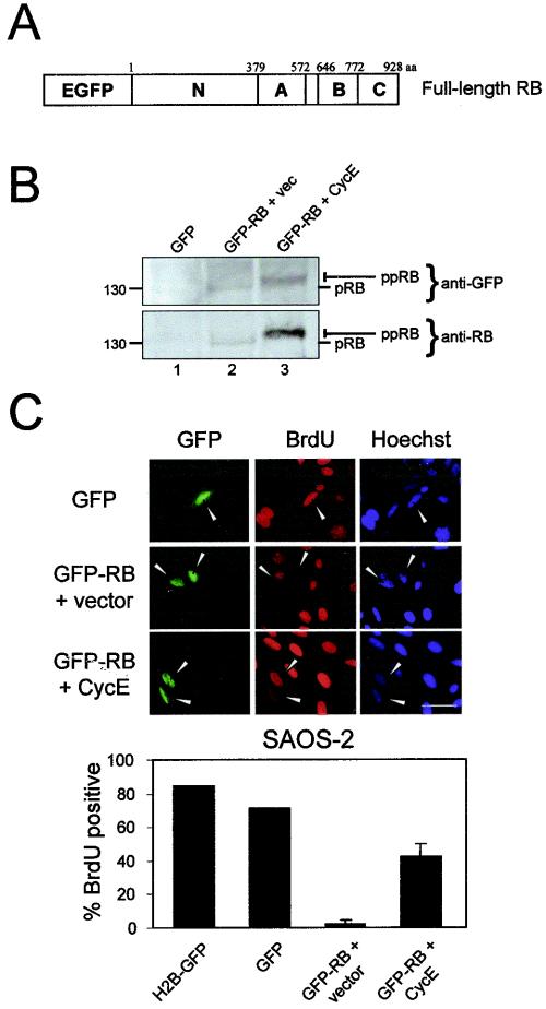
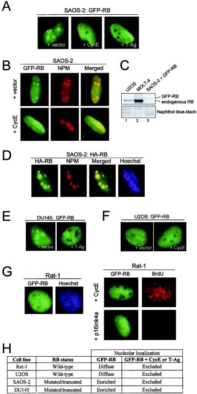
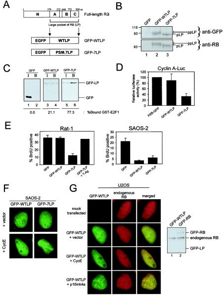
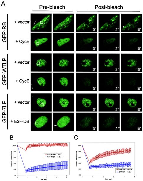
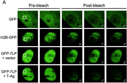
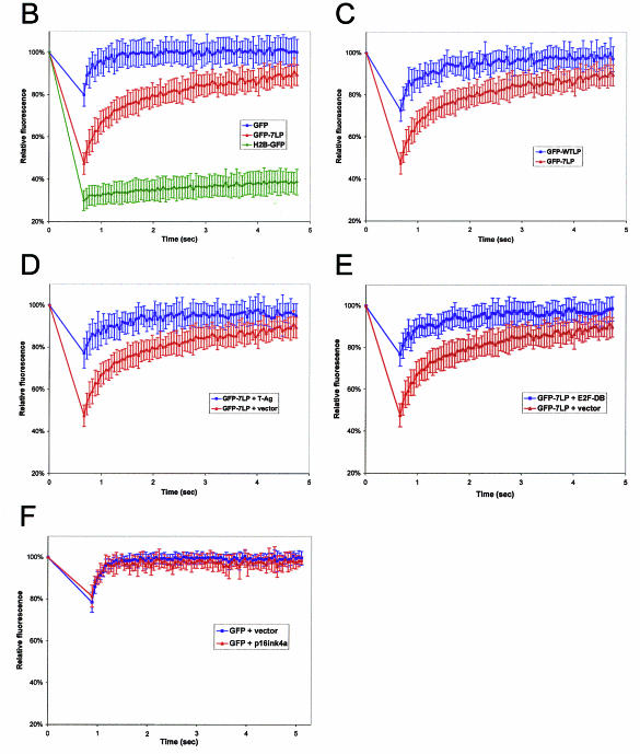
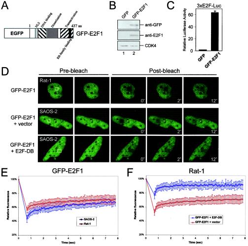
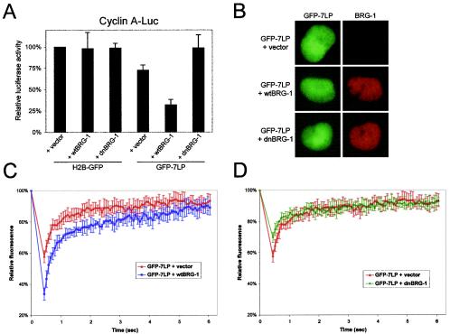
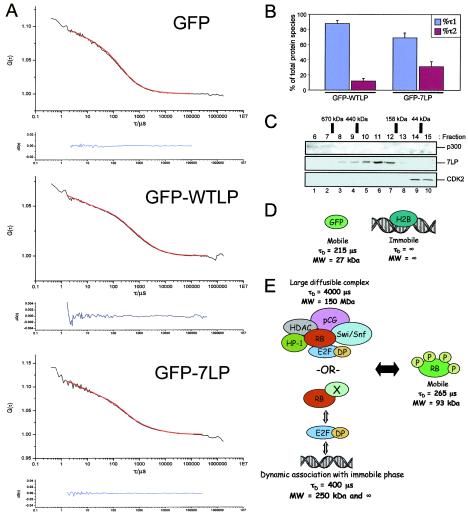
The retinoblastoma tumor suppressor, RB, assembles multiprotein complexes to mediate cell cycle inhibition. Although many RB binding partners have been suggested to underlie these functions, the validity of these interactions on the behavior of RB complexes in living cells has not been investigated. Here, we studied the dynamic behavior of RB by using green fluorescent protein-RB fusion proteins. Although these proteins were universally nuclear, phosphorylation or oncoprotein binding mediated their active exclusion from the nucleolus. In vivo imaging approaches revealed that RB exists in dynamic equilibrium between a highly mobile and a slower diffusing species, and genetic lesions associated with tumorigenesis increased the fraction of RB in a highly mobile state. The RB complexes dictating cell cycle arrest were surprisingly dynamic and harbored a relatively short residence time on chromatin. In contrast, this rapid exchange was attenuated in cells that are hypersensitive to RB, suggesting that responsiveness may inversely correlate with mobility. The stability of RB dynamics within the cell was additionally modified by the presence and function of critical corepressors. Last, the RB-assembled complexes present in living cells were primarily associated with E2F binding sites in chromatin. In contrast to RB, E2F1 consistently maintained a stable association with E2F sites regardless of cell type. Together, these results elucidate the kinetic framework of RB tumor suppressor action in transcriptional repression and cell cycle regulation.


Figures

FIG. 1.
FIG. 1.
GFP-RB fusion protein localizes to the nucleus and retains cell cycle-inhibitory activity. (A) Enhanced GFP was fused to the N terminus of wild-type RB. The N-terminal, A and B pocket, and C-terminal domains are indicated. aa, amino acids. (B) SAOS-2 cells were transfected with GFP (lane 1), GFP-RB and empty vector (vec) (lane 2), or GFP-RB and cyclin E (CycE) (lane 3). Immunoblotting was performed to detect either GFP or RB. The hyperphosphorylated (ppRB) or hypophosphorylated (pRB) bands are indicated on the right. Molecular masses (in kilodaltons) are indicated on the left. (C) SAOS-2 cells were transfected with GFP, GFP-RB and empty vector, or GFP-RB and cyclin E. (Upper panel) BrdU incorporation was detected by indirect immunofluorescent staining. Bar, 20 μm. (Lower panel) Quantitation of BrdU incorporation from two independent experiments with over 100 cells counted per experiment.
FIG. 2.
FIG. 2.
Hyperphosphorylated GFP-RB is excluded from the nucleolus. (A) SAOS-2 cells were transfected with GFP-RB and either vector, cyclin E (CycE), or SV40 T-Ag. (B) SAOS-2 cells as described for panel A were permeabilized, and immunofluorescent staining was performed with an antibody directed against NPM. GFP fluorescence, NPM staining, and merged images of nuclei are shown. (C) U2OS (lane 1), MOLT-4 (lane 2), and SAOS-2 cells transfected with GFP-RB (lane 3) were harvested, and whole-cell lysates were resolved by SDS-PAGE. RB was detected by immunoblotting. Naphthol blue-black staining is shown as a loading control. (D) SAOS-2 cells were transfected with HA-tagged RB. HA-RB was detected by indirect immunofluorescence. (E) DU145 cells were transfected with GFP-RB and either vector or T-Ag. (F) U2OS cells weretransfected with GFP-RB and either vector or cyclin E. (G) (Left panel) Rat-1 cells stably transfected with GFP-RB. (Right panel) Rat-1 cells stably expressing GFP-RB were infected with recombinant adenovirus encoding either p16ink4a or cyclin E for 16 h. BrdU incorporation was detected by indirect immunofluorescence. (H) Summary of GFP-RB localization in various cell lines. (A, B, and D to G) Representative photomicrographs taken at ×60 magnification.
FIG. 3.
FIG. 3.
The N-terminal domain of RB may contribute to nucleolar localization but is not required for cell cycle inhibition. (A) The critical growth inhibitory and tumor suppressive region of the retinoblastoma protein is comprised of an A and B pocket domain and the C terminus. These regions are termed the large pocket (LP) domain of RB. The cDNA encoding enhanced GFP (EGFP) was fused to wild-type LP (GFP-WTLP) and a phosphorylation site mutant LP (GFP-7LP). aa, amino acids. (B) C33A cells were transfected with either GFP (lane 1), GFP-WTLP (lane 2), or GFP-7LP (lane 3). Immunoblotting was performed to detect GFP (upper panel) or RB (lower panel). The hyperphosphorylated (ppLP) and hypophosphorylated (pLP) bands are indicated on the right. Molecular masses (in kilodaltons) are indicated on the left. (C) U2OS cells weretransfected with either GFP (lanes 1 and 2), GFP-WTLP (lanes 3 and 4), or GFP-7LP (lanes 4 and 6). Input (I) (lanes 1, 3, and 5) and GST-E2F1-bound (B) (lanes 2, 4, and 6) fractions were resolved by SDS-PAGE, and GFP was detected by immunoblotting. The percentage of GFP bound to E2F1 was determined by using Metamorph software. (D) Rat-1 cells were transfected with CMV-β-galactosidase, human −608Cyclin A-Luc reporter plasmid, and either H2B-GFP, GFP-WTLP, or GFP-7LP. Relative luciferase activity was determined and set to 100% for H2B-GFP. (E) Rat-1 cells (left panel) were transfected with either GFP, GFP-WTLP, or GFP-7LP and empty vector or GFP-7LP with SV40 T-Ag. SAOS-2 cells (right panel) were transfected with GFP, GFP-WTLP, or GFP-7LP. BrdU incorporation was determined by indirect immunofluorescence. Data shown are from over 200 cells counted. (F) SAOS-2 cells were transfected with GFP-WTLP or GFP-7LP and either empty vector or cyclin E. Representative photomicrographs were taken at a ×60 magnification. (G) (Left panel) U2OS cells were either mock transfected or transfected with GFP-WTLP and either vector, cyclin E (CycE), or p16ink4a. Endogenous RB was simultaneously detected by indirect immunofluorescence. Representative photomicrographs are shown at a magnification of ×54. (Right panel) U2OS cells transfected with either GFP-WTLP (lane 1) or GFP-RB (lane 2) were harvested, and whole-cell lysates were resolved by SDS-PAGE. Full-length RB, but not WTLP, was detected by immunoblotting with an N-terminal-specific antibody (G3-245).
FIG. 4.
FIG. 4.
FRAP analysis reveals immobility of active GFP-RB in SAOS-2 cells. (A) SAOS-2 cells were transfected with either GFP-RB, GFP-WTLP, or GFP-7LP and either empty vector, cyclin E, or E2F-DB as indicated. At 24 h posttransfection, cells were subjected to nuclear FRAP analysis. (B and C) Recovery curves from SAOS-2 cells shown in panel A. Relative fluorescence intensities were determined by comparing the fluorescence intensity of a distal unbleached region of the nucleus to the photobleached area, and the results are plotted over time. The white boxes indicate the bleached areas (2.9 by 2.9 μm2).
FIG. 5.
FIG. 5.
FRAP analysis uncovers dynamic regulation of GFP-LP by phosphorylation, oncoprotein binding, and E2F site availability. (A) Rat-1 cells were transfected as indicated. At 24 h posttransfection, cells were subjected to nuclear FRAP analysis. As a control, no recovery of fusion proteins was observed after bleaching of chemically fixed cells. The white boxes indicate the bleached areas (2.9 by 2.9 μm2). (B to F) Recovery curves, with relative fluorescence intensity plotted over time.
FIG. 5.
FIG. 5.
FRAP analysis uncovers dynamic regulation of GFP-LP by phosphorylation, oncoprotein binding, and E2F site availability. (A) Rat-1 cells were transfected as indicated. At 24 h posttransfection, cells were subjected to nuclear FRAP analysis. As a control, no recovery of fusion proteins was observed after bleaching of chemically fixed cells. The white boxes indicate the bleached areas (2.9 by 2.9 μm2). (B to F) Recovery curves, with relative fluorescence intensity plotted over time.
FIG. 6.
FIG. 6.
GFP-E2F1 dynamics are not cell type dependent and depend on E2F site occupancy. (A) Enhanced GFP (EGFP) was fused to the N terminus of human E2F1. The domain structure is indicated. aa, amino acids. (B) U2OS cells were transfected with GFP (lane 1) or GFP-E2F1 (lane 2), whole-cell extracts were resolved by SDS-PAGE, and the indicated proteins were detected by immunoblotting. (C) U2OS cells were transfected with CMV-β-galactosidase, 3XE2F-Luc reporter, and either GFP or GFP-E2F1. The relative luciferase activity was determined. The data shown are from two independent experiments. (D) Rat-1 or SAOS-2 cells were transfected with GFP-E2F1 alone or with either vector or E2F1-DB as indicated. At 24 h posttransfection, cells were subjected to nuclear FRAP analysis. The white boxes indicate the bleached areas (2.9 by 2.9 μm2). (E and F) Recovery curves for GFP-E2F1 from panel D are shown, with relative fluorescence intensity plotted over time.
FIG. 7.
FIG. 7.
GFP-7LP recruitment of functional BRG-1 forms an active repressor module with distinct dynamics. (A) C33A cells were transfected with CMV-β-galactosidase, cyclin A-Luc reporter, H2B-GFP, or GFP-7LP and either vector, wtBRG-1, or dnBRG-1 as indicated. The relative luciferase activity was determined. The data shown are from three independent experiments. (B) SW13 cells were transfected with GFP-7LP and either vector, wtBRG-1, or dnBRG-1. BRG-1 was detected by indirect immunofluorescence. Representative photomicrographs were taken at a magnification of ×60. (C and D) SW13 cells transfected as described for panel B were utilized for nuclear FRAP analysis. Relative fluorescence intensity is plotted over time.
FIG. 8.
FIG. 8.
FCS analysis of RB complex dynamics. (A) Rat-1 cells expressing either GFP, GFP-WTLP, or GFP-7LP were subjected to nuclear FCS analysis. Shown are mean autocorrelation curves (upper plots) derived from fluctuations of fluorescence intensity over time measured for 4-s intervals (lower plots). Autocorrelation curves were fit to a two-component model of free diffusion in two dimensions with Origin software to derive the translational diffusion time and number of molecules diffusing through the confocal volume with Dfast and Dslow. (B) The data obtained from FCS identified that GFP-LP diffused as two distinct species: a mobile species with rapid diffusion rate (τ1) and a 10- to 20-fold-more-slowly diffusing complex (τ2). The percentages of total protein species diffusing through the confocal volume with τ1 and τ2 are shown. (C) A2-4 cells expressing 7LP were harvested, whole-cell lysate was subjected to gel filtration, and eluted fractions were resolved by SDS-PAGE. The indicated proteins were detected by immunoblotting. Molecular mass standards are indicated. (D and E) Proposed models for the behavior of GFP, H2B, and RB in vivo. RB can potentially interact with multiple cellular proteins, forming a large diffusible complex. Data presented here support a model wherein RB and essential transcriptional corepressors dynamically exchange with E2F-binding sites in chromatin.

References

    1. Alcalay, M., L. Tomassoni, E. Colombo, S. Stoldt, F. Grignani, M. Fagioli, L. Szekely, K. Helin, and P. G. Pelicci. 1998. The promyelocytic leukemia gene product (PML) forms stable complexes with the retinoblastoma protein. Mol. Cell. Biol. 18:1084-1093. - PMC - PubMed
    1. Angus, S. P., A. F. Fribourg, M. P. Markey, S. L. Williams, H. F. Horn, J. DeGregori, T. F. Kowalik, K. Fukasawa, and E. S. Knudsen. 2002. Active RB elicits late G1/S inhibition. Exp. Cell Res. 276:201-213. - PubMed
    1. Angus, S. P., L. J. Wheeler, S. A. Ranmal, X. Zhang, M. P. Markey, C. K. Mathews, and E. S. Knudsen. 2002. Retinoblastoma tumor suppressor targets dNTP metabolism to regulate DNA replication. J. Biol. Chem. 277:44376-44384. - PubMed
    1. Chew, Y. P., M. Ellis, S. Wilkie, and S. Mittnacht. 1998. pRB phosphorylation mutants reveal role of pRB in regulating S phase completion by a mechanism independent of E2F. Oncogene 17:2177-2186. - PubMed
    1. Dahiya, A., S. Wong, S. Gonzalo, M. Gavin, and D. C. Dean. 2001. Linking the Rb and polycomb pathways. Mol. Cell 8:557-569. - PubMed

Publication types

MeSH terms

LinkOut - more resources