. 2003 Nov;23(22):8233-45.
doi: 10.1128/MCB.23.22.8233-8245.2003.

Neurons expressing the highest levels of gamma-synuclein are unaffected by targeted inactivation of the gene

Affiliations

Neurons expressing the highest levels of gamma-synuclein are unaffected by targeted inactivation of the gene

Natalia Ninkina et al. Mol Cell Biol. 2003 Nov.

Abstract

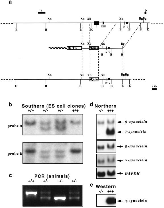
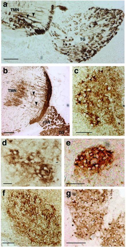
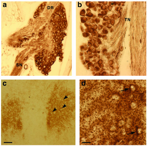
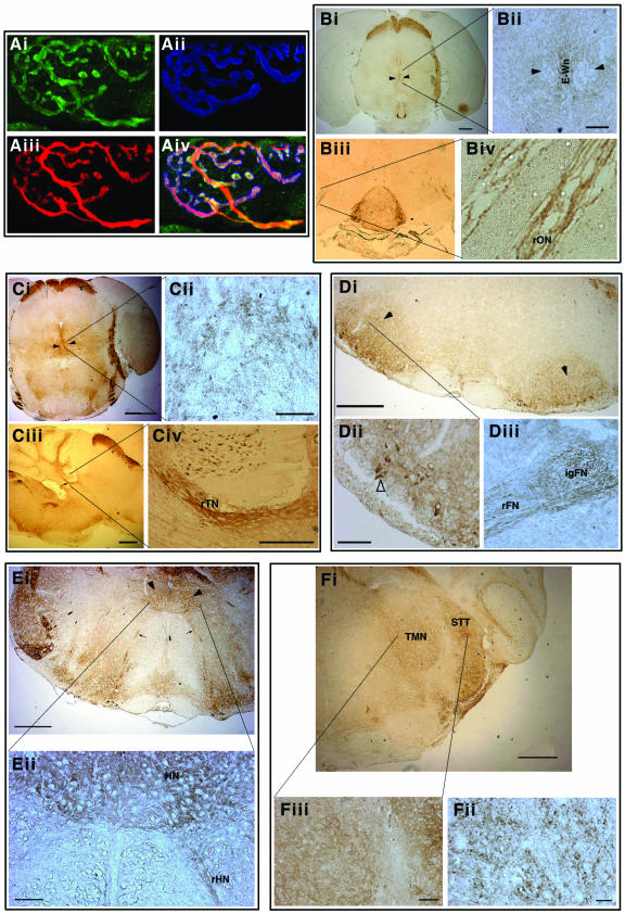
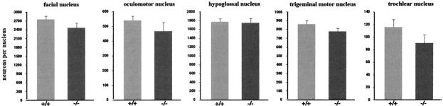
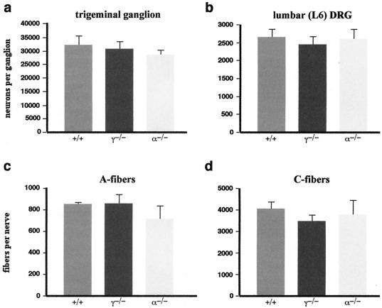
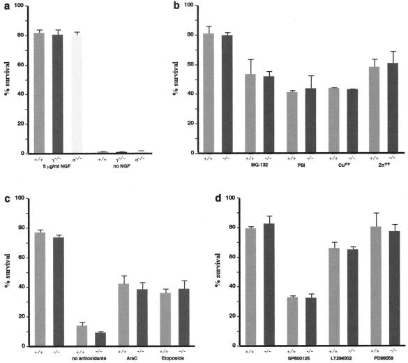
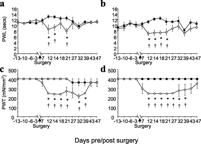
Homologous recombination in ES cells was employed to generate mice with targeted deletion of the first three exons of the gamma-synuclein gene. Complete inactivation of gene expression in null mutant mice was confirmed on the mRNA and protein levels. Null mutant mice are viable, are fertile, and do not display evident phenotypical abnormalities. The effects of gamma-synuclein deficiency on motor and peripheral sensory neurons were studied by various methods in vivo and in vitro. These two types of neurons were selected because they both express high levels of gamma-synuclein from the early stages of mouse embryonic development but later in the development they display different patterns of intracellular compartmentalization of the protein. We found no difference in the number of neurons between wild-type and null mutant animals in several brain stem motor nuclei, in lumbar dorsal root ganglia, and in the trigeminal ganglion. The survival of gamma-synuclein-deficient trigeminal neurons in various culture conditions was not different from that of wild-type neurons. There was no difference in the numbers of myelinated and nonmyelinated fibers in the saphenous nerves of these animals, and sensory reflex thresholds were also intact in gamma-synuclein null mutant mice. Nerve injury led to similar changes in sensory function in wild-type and mutant mice. Taken together, our data suggest that like alpha-synuclein, gamma-synuclein is dispensable for the development and function of the nervous system.


Figures

FIG. 1.
FIG. 1.
Targeted inactivation of the mouse γ-synuclein gene. (a) Scheme for deletion of exons I, II, and III and promoter region of the mouse γ-synuclein gene by homologous recombination. The organizations of the wild-type genomic locus (top), targeting vector (middle), and resulting knockout locus (bottom) are shown. Restriction endonuclease sites: E, EcoRI; B, BamHI; Xb, XbaI; K, KpnI; Bg, BglII. Hybridization probes a and b, which were used for the analysis of homologous recombination, are also shown. (b) Examples of analysis of homologous recombination in ES cell lines by Southern hybridization. DNAs from four neomycin-resistant ES cell lines were digested with EcoRI and hybridized with either probe a or probe b. Only a 20-kb wild-type band is revealed in two clones with random insertion of a PGK-neo cassette, and the homologous recombination in two other clones results in the appearance of a 17-kb band. Similar results were obtained when DNA was digested with BamHI and hybridized with probe c. (c) Example of PCR-based genotyping of mice from a litter of two heterozygous parents. (d) Expression of mRNAs encoding members of the synuclein family in the retinas of wild-type and γ-synuclein null mutant mice. Results of Northern hybridization with a full-length mouse γ-synuclein cDNA probe, a mouse β-synuclein-specific probe, a mouse α-synuclein-specific probe, and a GAPDH (glyceraldehyde-3-phosphate dehydrogenase) probe are shown. Note that under the hybridization and washing conditions used, the γ-synuclein cDNA probe cross-hybridized with β-synuclein transcript (upper panel). High-stringency washes completely eradicated this hybridization signal, with no effect on hybridization with γ-synuclein transcript. (e) A Western blot of 10 μg of total spinal cord proteins of wild-type and γ-synuclein null mutant mice was probed with mouse γ-synuclein-specific SK23 antibody.
FIG.2.
FIG.2.
γ-Synuclein in embryonic mouse sensory and motoneurons. Anti-γ-synuclein staining of E12 (a) and E15 (b) trigeminal motor nuclei and trigeminal ganglia and of E18 trigeminal motor nucleus (c), E18 oculomotor nucleus (d), E18 trochlear nucleus (e), E18 facial nucleus (f), and E18 hypoglossal nucleus (g) is shown. All nuclei contain labeled neuronal cell bodies, but dotted neuropil stain becoming also obvious in all ganglia at E18. Arrowheads show γ-synuclein-positive axons of E12 and E15 motoneurons of the trigeminal motor nucleus (TMN). Sensory neurons of the trigeminal ganglion (TG) are also intensively stained, as is the root of the trigeminal nerve (rTN). Bars, 100 μm (a, c, e, and f), 200 μm (b and g), and 50 μm (d).
FIG. 3.
FIG. 3.
γ-Synuclein in postnatal mouse sensory and motoneurons. γ-Synuclein is localized in the cytoplasm of cell bodies of mouse P2 DRG (a) and trigeminal ganglion (b) neurons as well as in nerve fibres in the dorsal root (DR), spinal nerve (SN), and trigeminal nerve (TN). In P2 oculomotor nucleus (c), only γ-synuclein-negative cell bodies (arrowheads) are seen on the background of neuropil staining, whereas in P2 trigeminal motor nucleus (d), cytoplasmic staining can be detected in some neurons (arrows). Bars, 100 μm (a and c) and 20 μm (b and d).
FIG.4.
FIG.4.
γ-Synuclein in adult mouse motoneurons. (A) γ-Synuclein in motor axons and nerve terminals at the neuromuscular synapse. Triple immunofluorescent staining of whole-mount preparations of mouse traingularis sterni muscle is shown. γ-Synuclein (Ai, green) is colocalized with neurofilaments in the axon and SV2 in presynaptic terminals (Aiii, red) but not with acetylcholine receptors on postsynaptic membrane (Aii, blue [stained with AlexaFluor 647-conjugated μ-bungarotoxin]). Three images are merged in panel Aiv. (B) γ-Synuclein in oculomotor nuclei (arrowheads) and the root of oculomotor nerve (rON). A higher magnification shows axonal staining in the nerve root (panel Biv), the absence of γ-synuclein in cell bodies of motoneurons, and positive staining of neuronal cell bodies in the median Edinger-Westphal nucleus (E-Wn, panel Bii). (C) γ-Synuclein in trochlear nuclei (arrowheads in panel Ci) and the root of trochlear nerve (arrow in panel Ciii and rTN in panel Civ). A higher magnification shows intense staining of motoneuron axons in the nerve (Civ) and only a dotted neuropil staining in the nucleus (Cii). (D) γ-Synuclein in facial nuclei (arrowheads) and the facial nerve. Axonal staining is evident in the internal genu (IgFN) and the root (rFN) of the nerve (panel Diii). A higher magnification reveals γ-synuclein in the cytoplasm of a few motoneurons (open arrowhead in panel Dii). (E) γ-Synuclein in hypoglossal nuclei (arrowheads in panel Ei and HN in panel Eii) and the root of hypoglossal nerve (rHN in panel Eii). A higher magnification shows positive staining of the nerve root and neuropil staining in the nucleus (Eii). (F) γ-Synuclein in the trigeminal motor nucleus (TMN) and the spinal trigeminal tract (STT). Neuronal cell bodies are not stained; only dotted neuropil staining is evident at the highest magnification (Fiii).
FIG. 5.
FIG. 5.
Average total numbers of motoneurons in brain stem motor nuclei of wild-type (+/+) and γ-synuclein null mutant (-/-) adult mice. Means and standard errors from data obtained from analysis of at least 10 nuclei for each genotype are shown. Statistical analysis (two-tailed, unpaired Student's t test) showed no significant difference in cell numbers between the two genotypes for all nuclei (P > 0.5).
FIG. 6.
FIG. 6.
Numbers of sensory neurons and nerve fibers in wild-type and null mutant mice. (a and b) Average total number of neurons in trigeminal ganglia (a) and L6 lumbar DRG (b) of wild-type (+/+), γ-synuclein null mutant (γ−/−), and α-synuclein null mutant (α−/−) P2 mice. Means and standard errors of data obtained from analysis of at least 10 ganglia for each genotype are shown. Statistical analysis (Kruskal-Wallis one-way analysis of variance) showed no significant difference in cell numbers between all three genotypes for both ganglia (P > 0.6). (c and d) Average total numbers of myelinated A-fibers (c) and unmyelinated C-fibers (d) in adult mouse saphenous nerves. Means and standard errors of data obtained from analysis of at least five nerves for each genotype are shown. Statistical analysis (Kruskal-Wallis one-way analysis of variance) showed no significant difference in numbers for both types of fibers between all three genotypes (P > 0.4).
FIG. 7.
FIG. 7.
Survival of P2 mouse trigeminal ganglion neurons in dissociated primary culture. Cultures were prepared and treated with drugs as described in Materials and Methods. Bar charts illustrate survival of neurons 48 h after initial count and addition of drugs. The number of surviving neurons is expressed as a percentage of the initial count. Means and standard errors of data obtained from analysis of at least six culture dishes for each genotype in two independent experiments are shown. (a) Both γ-synuclein-deficient (γ−/−) and α-synuclein-deficient (α−/−) neurons have the same survival rate as wild-type (+/+) neurons in the presence of nerve growth factor (NGF) and are unable to survive in its absence (P > 0.8, Kruskal-Wallis one-way ANOVA). (b to d) Various treatments have the same effect on γ-synuclein deficient (−/−) neurons as they have on wild-type (+/+) neurons (P > 0.5 for all conditions; two-tailed, unpaired Student's t test). Proteasome inhibitors (5 μM MG-132 or 10 μM proteasome inhibitor I [PSI]) or heavy metal ions (30 μM CuSO4 or 75 μM ZnSO4) were added to neurons after the initial count at 24 h after plating (b). In one set of experiments neurons were plated in neurobasal medium supplemented with B27 without antioxidants (c). In other cases, DNA-damaging agents (10 μM cytosine arabinoside [AraC] or 10 μM Etoposide) or inhibitors of the JNK signaling pathway (20 μM SP600125), ERK signaling pathway (20 μM PD98059), or phosphatidylinositol 3-kinase signaling pathway (20 μM LY294002) were added to neurons after the initial count at 3 h after plating (c and d).
FIG. 8.
FIG. 8.
Behavioral analysis of mice with CCI to the sciatic nerve. Data show mean (± standard error of the mean) responses taken over a period of up to 13 days before and every 2 to 6 days following surgery in wild-type mice (a and c) (n = 9) and γ-synuclein null mutant mice (b and d) (n = 6). (a and b) Paw withdrawal latency (PWL) from a noxious thermal stimulus (Hargreaves' thermal stimulator) ipsilateral to CCI (○) showed significant differences between postoperative and preoperative values (†, P < 0.05; Kruskal-Wallis one-way analysis of variance) and from postoperative, contralateral (▪) values (*, P < 0.05 by Student's t test) for both wild-type (a) and mutant (b) mice. No thermal hyperalgesia was seen on the contralateral side. (c and d) Paw withdrawal thresholds (PWT) from mechanical stimulation (von Frey filaments) showed significant differences between postoperative and preoperative values on the side ipsilateral (○) to CCI (†, P < 0.05; Dunn's method analysis of variance on ranks) and between postoperative ipsilateral and contralateral (▪) values (*, P < 0.05, Mann-Whitney U-test) for both wild-type (c) and mutant (d) mice.

Similar articles

Cited by

References

    1. Abeliovich, A., Y. Schmitz, I. Farinas, D. Choi-Lundberg, W. H. Ho, P. E. Castillo, N. Shinsky, J. M. Verdugo, M. Armanini, A. Ryan, M. Hynes, H. Phillips, D. Sulzer, and A. Rosenthal. 2000. Mice lacking alpha-synuclein display functional deficits in the nigrostriatal dopamine system. Neuron 25:239-252. - PubMed
    1. Alimova-Kost, M. V., N. N. Ninkina, S. Imreh, N. V. Gnuchev, J. Adu, A. M. Davies, and V. L. Buchman. 1999. Genomic structure and chromosomal localization of the mouse persyn gene. Genomics 56:224-227. - PubMed
    1. Bennett, G. L., and Y.-K. Xie. 1988. A peripheral mononeuropathy in rat that produces disorders of pain sensation like those seen in man. Pain 33:87-109. - PubMed
    1. Biere, A. L., S. J. Wood, J. Wypych, S. Steavenson, Y. Jiang, D. Anafi, F. W. Jacobsen, M. A. Jarosinski, G. M. Wu, J. C. Louis, F. Martin, L. O. Narhi, and M. Citron. 2000. Parkinson's disease-associated alpha-synuclein is more fibrillogenic than beta- and gamma-synuclein and cannot cross-seed its homologs. J. Biol. Chem. 275:34574-34579. - PubMed
    1. Blackburn-Munro, G., and S. M. Fleetwood-Walker. 1999. The sodium channel auxiliary subunits beta1 and beta2 are differentially expressed in the spinal cord of neuropathic rats. Neuroscience 90:153-164. - PubMed

Publication types

MeSH terms