. 2003 Nov;23(22):8255-71.
doi: 10.1128/MCB.23.22.8255-8271.2003.

Female lethality and apoptosis of spermatocytes in mice lacking the UBR2 ubiquitin ligase of the N-end rule pathway

Affiliations

Female lethality and apoptosis of spermatocytes in mice lacking the UBR2 ubiquitin ligase of the N-end rule pathway

Yong Tae Kwon et al. Mol Cell Biol. 2003 Nov.

Abstract

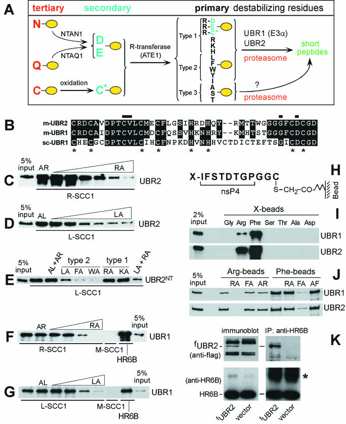
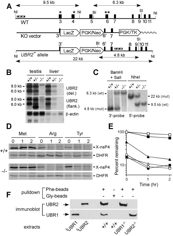
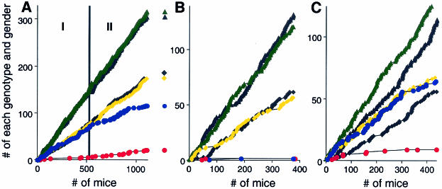
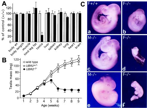
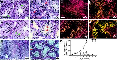
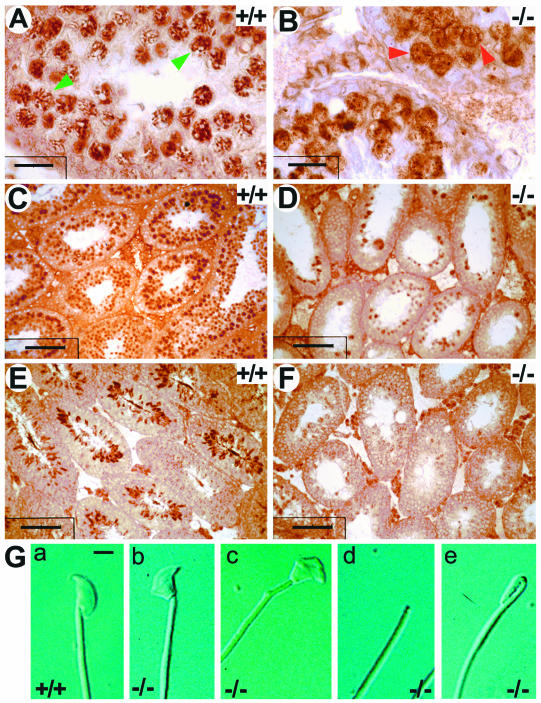
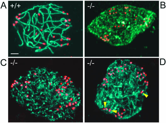
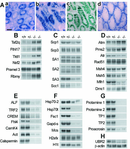
Substrates of the ubiquitin-dependent N-end rule pathway include proteins with destabilizing N-terminal residues. UBR1(-/-) mice, which lacked the pathway's ubiquitin ligase E3alpha, were viable and retained the N-end rule pathway. The present work describes the identification and analysis of mouse UBR2, a homolog of UBR1. We demonstrate that the substrate-binding properties of UBR2 are highly similar to those of UBR1, identifying UBR2 as the second E3 of the mammalian N-end rule pathway. UBR2(-/-) mouse strains were constructed, and their viability was found to be dependent on both gender and genetic background. In the strain 129 (inbred) background, the UBR2(-/-) genotype was lethal to most embryos of either gender. In the 129/B6 (mixed) background, most UBR2(-/-) females died as embryos, whereas UBR2(-/-) males were viable but infertile, owing to the postnatal degeneration of the testes. The gross architecture of UBR2(-/-) testes was normal and spermatogonia were intact as well, but UBR2(-/-) spermatocytes were arrested between leptotene/zygotene and pachytene and died through apoptosis. A conspicuous defect of UBR2(-/-) spermatocytes was the absence of intact synaptonemal complexes. We conclude that the UBR2 ubiquitin ligase and, hence, the N-end rule pathway are required for male meiosis and spermatogenesis and for an essential aspect of female embryonic development.


Figures

FIG.1.
FIG.1.
Mouse UBR2 is the ubiquitin ligase of the N-end rule pathway. (A) N-end rule pathway in mammals. N-terminal residues are indicated by single-letter abbreviations for amino acids. The yellow ovals designate the rest of a protein substrate. (B) Alignment of the sequences of the UBHC (Ub, His, and Cys) domain (12, 25) of m-UBR2 (residues 112 to 153), m-UBR1, and sc-UBR1. The asterisks indicate conserved Cys and His. Horizontal bars indicate residues important for the binding of type 1 destabilizing N-terminal residues by sc-UBR1 (see Results). (C to G) GST pull-down assays. Equal amounts of an extract from S. cerevisiae containing either m-fUBR1 (E3α), m-fUBR2, or its 1,041-residue N-terminal fragment (m-fUBR21-1041) were incubated with glutathione-Sepharose beads preloaded with X-SCC1-GST (X = Arg [R], Leu [L], or Met [M]) or with GST-HR6B. The incubations were performed in the presence or absence of the indicated dipeptides: RA, Arg-Leu; AR, Ala-Arg; LA, Leu-Ala; AL, Ala-Leu; FA, Phe-Ala; WA, Trp-Ala; KA, Lys-Ala. The bound proteins were eluted, fractionated by SDS-PAGE, and immunoblotted with anti-Flag antibody. The 5% input lanes refer to a directly loaded sample of yeast extract that corresponded to 5% of the amount of extract used in GST assays. The concentrations of a competitor peptide were 1 μM, 10 μM, 0.1 mM, 0.5 mM, 1 mM, and 2 mM for the results shown in panels C and D, and 1 mM in for the results shown in panels E, F, and G. (H to K) Pull-down assay with peptide-linked microbeads. (H) Bead-linked 12-mer peptides. X = Arg, Phe, Gly, Ser, Thr, Ala, or Asp. (I) m-UBR1 and m-UBR2 bind to Arg (type 1) and Phe (type 2) destabilizing N-terminal residues but not to the other tested N-terminal residues. (J) Binding competition assays with m-UBR1 and m-UBR2, the 12-mer peptides bearing N-terminal Arg or Phe, and competitor dipeptides. (K) m-UBR2 binds to the HR6B E2 enzyme. Extracts (0.1 mg of protein) from control NIH 3T3 cells and NIH 3T3 cells stably expressing m-fUBR2 from the PCMV promoter were fractionated by SDS-12% PAGE (left panel) followed by immunoblotting with anti-Flag (top panel) or anti-HR6B antibody (bottom panel). Extracts from the same cell lines (1 mg of protein) were immunoprecipitated with anti-HR6B (right panel) followed by SDS-12% PAGE and immunoblotting with anti-Flag (top panel) or anti-HR6B antibody (bottom panel). The asterisk designates the band of light chain immunoglobulin G.
FIG. 2.
FIG. 2.
Construction of UBR2−/− mouse strains. (A) Targeting vector. A map of the ∼30-kb 5′-proximal region of the ∼96-kb m-UBR2 gene, the targeting vector, and the deletion disruption UBR2−/− allele. Exons are designated by vertical rectangles. Solid and wavy horizontal lines designate, respectively, the mouse and plasmid DNAs. The directions of gene transcription are indicated as well. Exons 3 and 4, marked by single asterisks, and exon 6, marked by a double asterisk, contain conserved residues that are essential for integrity of the substrate-binding sites of sc-UBR1. NI, NheI; BI, BamHI; SI, SalI. Southern hybridization probes are indicated by striped rectangles. PGK, phosphatidylglycerol kinase; TK,thymidine kinase. (B) Northern analysis of m-UBR2 expression with total RNA from the testes and livers of adult +/+, UBR+/−, and UBR2−/− mice. The probes used were either the 350-bp UBR2 cDNA fragment (nt 383 to 731) that was deleted in the UBR2−/− allele (data not shown) or the 2.3-kb UBR2 cDNA fragment (nt 863 to 3227) adjacent to the deleted region (upper panel) or the human β-actin cDNA fragment (lower panel). The wild-type 8.0- and 6.0-kb UBR2 mRNAs are indicated on the left. (C) Southern analysis of BamHI/SalI-cut (3′ probe) and NheI-cut (5′ probe) mouse tail DNA. The 760-bp BamHI/SalI 3′ probe detected 6.3- and 4.8-kb UBR2 fragments for the wild-type (wt) and mutant (mut) UBR2 alleles, respectively. The 1.2-kb NheI 5′ probe detected 9.5-kb (wild type) and 22-kb (mutant) fragments. (D) Pulse-chase analysis with +/+ and UBR2−/− EF cell lines. EFs were transfected with plasmids expressing fDHFRh-UbR48-X-nsP4f, which yielded the fDHFRh-UbR48 reference protein (designated DHFR), and an X-nsP4f (X-nsP4-Flag) test protein (X = Met, Arg, or Tyr) (designated X-nsP4). Cells were labeled for 10 min with [35S]methionine and chased for 1 and 2 h. (E) Quantitation of the patterns in shown in panel D with PhosphorImager. For each time point, the ratio of 35S in X-nsP4f to 35S in the Met-DHFR-UbR48 reference protein at the same time point was plotted as the percentage of that ratio relative to that for Met-nsP4f (which bore a stabilizing N-terminal residue) at time zero (the beginning of chase). Open and closed symbols designate the results with +/+ and UBR2−/− EFs, respectively. Squares, Met-nsP4f; circles, Arg-nsP4f; triangles, Tyr-nsP4f. (F) Extracts from +/+, UBR1−/−, and UBR2−/− EFs were assayed (see Results) by using peptide pull-down assays and immunoblotting with anti-UBR1 and anti-UBR2 antibodies. fUBR1 and fUBR2 designate S. cerevisiae extracts expressing Flag-tagged m-UBR1 and m-UBR2.
FIG. 3.
FIG. 3.
Viability and other phenotypes of UBR2−/− mice depend on gender and genetic background. (A) Tabulation of progeny from matings between UBR2+/− mice of the 129SvImJ/(C57BL/6J) hybrid background, designated 129/B6 below. Green and black triangles, UBR2+/− males and females, respectively; yellow and black squares, +/+ males and females, respectively; blue and red circles, UBR2−/− males and females, respectively. The numbers on the abscissa refer to 1,106 offspring of UBR2+/− intercrosses. These numbers are temporally ordered, in that the tabulation of mice produced from earlier UBR2+/− intercrosses precede, on the abscissa, the tabulation of mice produced from later intercrosses. The accumulated numbers of intercross progeny of each genotype (+/+, UBR2+/−, or UBR2−/−) of a given gender are plotted on the ordinate. To produce 129/B6 UBR2−/− mice, the founder chimeras were mated with +/+ C57BL/6 females, yielding UBR2+/− 129/B6 mice followed by intercrosses. The aging breeding pairs of initial UBR2+/− mice were replaced, in the course of these experiments, with F2 generation UBR2+/− 129SvJ/(C57BL/6J) (129/B6) mice. For the actual numbers of progeny with specific genotypes, see Table 1. Phase I and phase II are described in Results. (B) Same as in panel A, except that the offspring were from UBR2+/− intercrosses in the 129SvImJ (inbred) background. (C) Same as in panel A, except that the offspring were from UBR2+/− intercrosses in the 129SvJ/CD1 background.
FIG. 4.
FIG. 4.
Testis degeneration and female lethality in UBR2−/− mice. (A) Weights of organs and tissues in UBR2−/− male mice [129SvJ/(C57BL/6J)] expressed as percentages of the corresponding weights for age-matched +/+ mice. The comparison involved 24 pairs of 2-month-old male mice (13 +/+, 11 UBR2+/−, and 24 UBR2−/−) produced through UBR2+/− intercrosses. Muscle, hind leg muscle; hind leg fat, fat pad of the hind leg. (B) Testis weights of +/+, UBR2+/−, and UBR2−/− mice [129SvJ/(C57BL/6J)] as a function of postnatal age. The total body weights of UBR2−/− mice were approximately equal to those of their +/+ littermates. (C) Whole-mount views of unstained +/+ and UBR2−/− embryos of the 129SvJ/(C57BL/6J) background. (a and b) E9.5 +/+ female embryo (a) and its UBR2−/− female littermate (b). (c and d) E9.5 UBR2−/− male embryo (c) and its UBR2−/− female littermate (d). (e and f) E11.5 UBR2−/− male embryo (e) and its UBR2−/− female littermate (f).
FIG. 5.
FIG. 5.
Arrest and apoptosis of spermatocytes in UBR2−/− mice. (A to F) Hematoxylin-eosin staining of testis sections from +/+ mice (A, C, and E) and UBR2−/− littermates (B, D, and F) of the 129SvJ/(C57BL/6J) background at 2 weeks (A and B), 3 weeks (C and D), and 8 weeks (E and F). Green arrowheads in panels A and C, pachytene spermatocytes; green arrow in panel C, round spermatid; red arrowheads in panels B, D, and F, apparently arrested spermatocytes; red arrow in panel D, aberrant pachytene spermatocyte. (G and H) Fluorescent TUNEL staining (and counterstaining with propidium iodide) of testis sections from 5-week-old +/+ male mice (G) and UBR2−/− littermates (H). (I and J) Same as panels G and H, respectively, but with 8-week-old littermates. (K) Quantitation of apoptosis in +/+ (○), UBR2+/− (▵), and UBR2−/− (•) testes as a function of postnatal age. The plotted numbers of apoptotic cells per 100 seminiferous tubules were derived from the examination of ∼400 tubule sections. Scale bars, 20 μm (A to D and G to J) and 60 μm (E and F).
FIG. 6.
FIG. 6.
Perturbed spermiogenesis in UBR2−/− mice. (A and B) Testis sections of 2-week-old +/+ (A) and UBR2−/− (B) littermates stained with anti-SCP3 antibody. Green arrowheads in panel A, typical pachytene spermatocytes; red arrowheads in panel B, arrested UBR2−/− spermatocytes. (C to F) Testis sections from 4-week-old +/+ (C and E) and UBR2−/− (D and F) littermates were stained with anti-tH2B (C and D) and anti-TP2 (E and F) antibodies. Scale bars, 20 μm (A and B), 7 μm (C), and 60 μm (D to F). (G) Appearance of +/+ (a) and UBR2−/− (b to e) spermatozoa.
FIG. 7.
FIG. 7.
Failure to assemble SC in UBR2−/− spermatocytes. Spermatocytes prepared from +/+ (A) and UBR2−/− (B to D) mice were analyzed by confocal microscopy, with antibody to SCP3 used to label the SC (green) and CREST antibody used to label centromeres (red). Yellow arrowheads, incompletely paired centromeres. Scale bar, 5 μm.
FIG. 8.
FIG. 8.
Northern and in situ hybridization analyses of UBR2−/− testes. (A) Distribution of lacZ-containing transcripts, derived from the UBR2−/− allele, in testis. Sections of 3-week-old testes from UBR2+/− (a) and UBR2−/− (b) mice were hybridized with antisense RNA probes specific for lacZ, which marked the deletion and/or disruption of the UBR2 allele. A UBR2-specific probe yielded a weak signal, hence the use of the lacZ-specific probe. No significant hybridization was detected with sections from these testes with a sense RNA probe (data not shown). (c) Testis sections from 3-week-old ATE1+/− mice which were heterozygous for the NLS-lacZ-marked deletion-disruption ATE1 allele (24) probed for lacZ expression as in panel a. (d) Distribution of UBR1 RNA in a 6-week-old UBR2+/− testis. Scale bar, 60 μm. (B to H) Northern analysis of RNA from +/+, UBR2+/−, and UBR2−/− testes of 6-week-old mice, with cDNA probes for the indicated genes.

References

    1. Alfonso, P. J., and W. S. Kistler. 1993. Immunohistochemical localization of spermatid nuclear transition protein 2 in the testes of rats and mice. Biol. Reprod. 48:522-529. - PubMed
    1. Ausubel, F. M., R. Brent, R. E. Kingston, D. D. Moore, J. A. Smith, J. G. Seidman, and K. Struhl (ed.). 2002. Current protocols in molecular biology. Wiley-Interscience, New York, N.Y.
    1. Baarends, W. M., E. Wassenaar, J. W. Hoogerbrugge, G. van Cappelen, H. P. Roest, J. Vreeburg, M. Ooms, J. H. J. Hoeijmakers, and J. A. Grootegoed. 2003. Loss of HR6B ubiquitin-conjugating activity results in damaged synaptonemal complex structure and increased crossing-over frequency during the male meiosis prophase. Mol. Cell. Biol. 23:1151-1162. - PMC - PubMed
    1. Baudat, F., K. Manova, J. P. Yuen, M. Jasin, and S. Keeney. 2000. Chromosome synapsis defects and sexually dimorphic meiotic progression in mice lacking Spo11. Mol. Cell 6:989-998. - PubMed
    1. Buonomo, S. B. C., R. K. Clyne, J. Fuchs, J. Loidl, F. Uhlmann, and K. Nasmyth. 2000. Disjunction of homologous chromosomes in meiosis I depends on proteolytic cleavage of the meiotic cohesin Rec8 by separin. Cell 103:387-398. - PubMed

Publication types

MeSH terms

Substances