. 2003 Nov;23(22):8352-62.
doi: 10.1128/MCB.23.22.8352-8362.2003.

The papillomavirus E8-E2C protein represses DNA replication from extrachromosomal origins

Affiliations

The papillomavirus E8-E2C protein represses DNA replication from extrachromosomal origins

Thomas Zobel et al. Mol Cell Biol. 2003 Nov.

Abstract

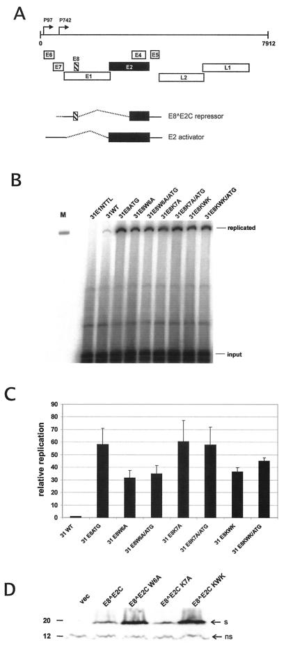
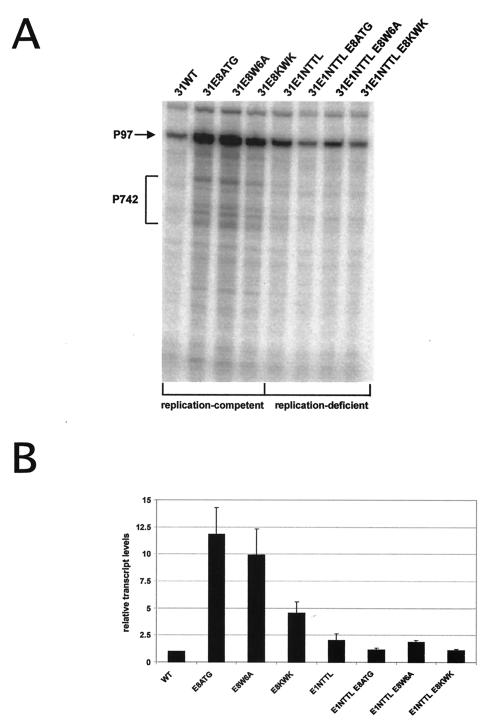
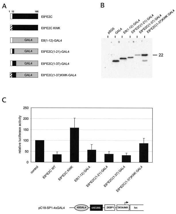
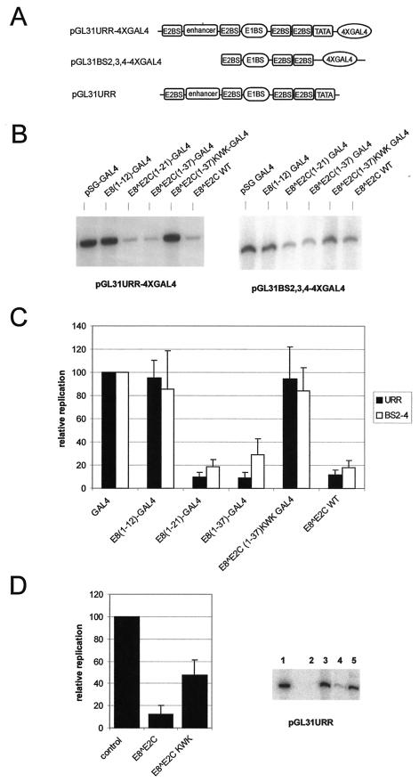
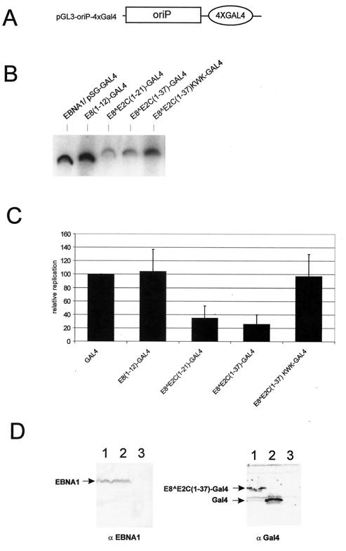
Carcinogenic DNA viruses such as high-risk human papillomaviruses (HPV) and Epstein-Barr-Virus (EBV) replicate during persistent infections as low-copy-number plasmids. EBV DNA replication is restricted by host cell replication licensing mechanisms. In contrast, copy number control of HPV genomes is not under cellular control but involves the viral sequence-specific DNA-binding E2 activator and E8-E2C repressor proteins. Analysis of HPV31 mutant genomes revealed that residues outside of the DNA-binding/dimerization domain of E8-E2C limit viral DNA replication, indicating that binding site competition or heterodimerization among E2 and E8-E2C proteins does not contribute to copy number control. Domain swap experiments demonstrated that the amino-terminal 21 amino acids of E8-E2C represent a novel, transferable DNA replication repressor domain, whose activity requires conserved lysine and tryptophan residues. Furthermore, E8-E2C (1-21)-GAL4 fusion proteins inhibited the replication of the plasmid origin of replication of EBV, suggesting that E8-E2C functions as a general replication repressor of extrachromosomal origins. This finding could be important for the development of novel therapies against persistent DNA tumor virus infections.


Figures

FIG. 1.
FIG. 1.
Replication properties of HPV31 E8 mutant genomes. (A) The linearized genome of HPV31 (nt 1 to 7912) with the various ORFs (E1 to E8, L1, and L2) and the positions of the major early promoter P97 and the major late promoter P742 are indicated by arrows on top. The E8 and E2 ORFs are highlighted by a striped box and black box, respectively. The structures of the spliced transcripts used to generate the E8Ê2C fusion protein and the full-length E2 protein are shown below. The carboxy terminus, which is part of both proteins, is responsible for dimerization among E2 proteins and sequence-specific DNA recognition (22). (B) Representative Southern blot of a transient replication analysis of HPV31 wild-type (31WT) and mutant genomes. The replication-deficient HPV31-E1NTTL genomeserved as a negative control. The positions of DpnI-resistant (replicated) DNA and DpnI-sensitive (input) DNA are indicated. Signal intensities of input DNA reveal similar transfection and recovery efficiencies for all constructs. One hundred picograms of EcoRI-digested HPV31 DNA was used as a size marker in lane M. (C) Graphic representation of the relative replication levels of HPV31 genomes. Signal intensities of DpnI-resistant DNA were quantified by phosphorimager analysis. The replication levels of the different HPV31 genomes are presented relative to that of wild-type HPV31 (31WT), which was set to 1. Error bars indicate standard deviations derived from data from three to five experiments. (D) Western blot analysis of E8Ê2C wild-type and mutant proteins. 293 cells were transfected with the empty expression vector pSG5 (vec) or expression vectors encoding wild-type E8E2C or mutant E8E2C W6A, K7A, or KWK proteins. A polyclonal antipeptide rabbit antiserum was used for the detection of E8E2C proteins. Bands representing E8E2C proteins are indicated by an arrow labeled “s.” A nonspecific band is also indicated (ns) and served as a loading control. A molecular mass marker (in kilodaltons) is shown on the left.
FIG. 2.
FIG. 2.
Analysis of viral transcripts from replication-competent and -deficient HPV31 genomes. (A) Representative RPA of mRNA isolated from normal human keratinocytes transiently transfected with different HPV31 genomes. The antisense probe spans HPV31 nt 678 to 919. The positions of transcripts initiated at the major early promoter P97 and spliced at the splice donor site at nt 877 or initiated at the major late promoter P742 are indicated by an arrow and a bracket, respectively. (B) Graphic representation of relative transcript levels initiated at P97 and spliced at nt 877. Transcript levels were quantitated by phosphorimager analysis. Levels are presented relative to HPV31 wild-type (WT) transfected cells, which were set to 1. Error bars indicate standard deviations derived from three independent experiments.
FIG. 3.
FIG. 3.
E8E2C-GAL4 fusion proteins act as transcriptional repressors. (A) Schematic representation of the structure of HPV31 E8E2C and E8E2C-GAL4 fusion proteins. The E8 part is depicted in white, the E2C part is depicted in black, and the GAL4 DBD is depicted in gray. The LDTR-deficient E8E2C KWK mutant protein was generated by changing residues 5 to 8 from KWK to AEA, and this is indicated by a striped box. Identical mutations were introduced into E8E2C(1-37) KWK-GAL4. Either 12, 21, or 37 amino-terminal residues of E8E2C or E8E2C KWK were fused to aa 1 to 147 of the DBD of the yeast transcription factor GAL4. (B) Western blot analysis of extracts isolated from transiently transfected 293 cells. Cells were transfected with the empty expression vector pSG5 or with plasmids expressing unfused or E8E2C-GAL4 proteins and analyzed with an antibody specific for the GAL4 DBD. A molecular mass marker is indicated to the right in kilodaltons. (C) SCC13 cells were cotransfected with 30 ng of expression vectors for E8E2C or E8E2C KWK, E8(1-12)-GAL4, E8E2C(1-21)-GAL4, E8E2C(1-37)-GAL4, E8E2C(1-37) KWK-GAL4, and 200 ng of the pC18-SP1-4xGAL4-luc luciferase reporter plasmid, respectively. In addition, 30 ng of pSG5 was included when transfecting expression vectors for GAL4 fusions, and 30 ng of pSG-GAL4 was added when E8E2C expression vectors were used. The average relative luciferase activities were calculated with respect to the activity of each construct in the presence of the parental pSG5 expression vector, which was set to 1. The luciferase activities are presented relative to the activity of pC18-SP1-4xGAL4-luc cotransfected withboth 30 ng of pSG5 and 30 ng of pSG-GAL4 expression vectors, which was set to 1 (control). Standard deviations are indicated by error bars. A Student′s t test analysis revealed that the observed differences between the control and E8(1-12)-GAL4 as well as between E8E2C(1-37)-GAL4 and E8E2C(1-37) KWK-GAL4 are statistically significant (P < 0.005). The structure of the promoter region of the luciferase reporter plasmid pC18-SP1-4xGAL4-luc is shown below the graph. Transcriptional control elements representing four GAL4 binding sites (4xGAL4), four E2BS (4xE2BS), two SP1 binding sites (2xSP1), the adenovirus major late promoter TATA-box/initiator element (TATA/Inr), and the RNA initiation site for the luciferase (luc) RNA are indicated.
FIG.4.
FIG.4.
E8E2C-GAL4 fusion proteins inhibit replication of the HPV31 origin. (A) Schematic structure of the replication origins of the different replication reporter plasmids. Binding sites for GAL4 (4xGAL4), E2 (E2BS), E1 (E1BS), and TATA-box binding (TATA) proteins, as well as the enhancer region of HPV31, which is composed of binding sites for cellular transcription factors, are shown. (B) Representative Southern blot analysis of the transient replication of the HPV31 origin of replication. SCC13 cells were transfected with either reporter plasmid pGL31URR-4xGAL4 (URR) or pGL31BS2,3,4-4xGAL4 (BS2-4) and expression vectors for HPV31 E1 and E2. In addition, cells received expression vectors for the GAL4 DBD (GAL4), the different E8E2C-GAL4 fusions, or wild-type E8E2C. (C) The levels of replicated reporter plasmids were analyzed by Southern blot analysis and quantitated by phosphorimager analysis. The replication levels of the different plasmid combinations are presented relative to the replication level of the origin reporter plasmids in the presence of E1, E2, and GAL4, which was set to 1. Error bars indicate standard deviations obtained from six independent experiments. A Student′s t test analysis revealed that the observed differences between E8(1-12)-GAL4 and E8E2C(1-21)-GAL4 as well as between E8E2C(1-37)-GAL4 and E8E2C(1-37) KWK-GAL4 are statistically significant (P < 0.005). (D) Southern blot analysis of the transient replication of the HPV31 origin in SCC13 cells. A representative Southern blot is shown on the right. Cells were either transfected with the reporter plasmid pGL31URR alone (lane 2) or together with expression vectors for HPV31 E1 and E2 (lanes 3 to 5). In addition, cells received expression vectors (300 ng) for wild-type E8E2C (lane 4) or the E8E2C KWK mutant (lane 5). Lane 1 received 100 pg of linearized pGLURR31 plasmid and served as a size marker. Data were quantitated by phosphorimager analysis and are presented in the graph on the right. Replication levels are presented relative to the HPV31 origin replication in the presence of E1 and E2 alone (control). Error bars indicate standard deviations obtained from seven independent experiments. Student′s t test analysis revealed that the difference between E8E2C and E8E2C KWK is statistically significant (P < 0.005).
FIG. 5.
FIG. 5.
E8E2C-GAL4 fusion proteins inhibit replication of the EBV oriP. (A) Schematic structure of the origin region of the replication reporter plasmid pGL3-oriP-4xGAL4. It consists of the oriP region from EBV, which is composed of multiple binding sites for EBNA1, and four binding sites for GAL4 (4xGAL4). (B) HeLa cells were transfected with the reporter plasmid pGL3-oriP-4xGAL4 and an expression vector for EBNA1. In addition, cells received expression vectors encoding the GAL4 DBD (pSG-GAL4), or the different E8-GAL4 fusions. The levels of DpnI-resistant (replicated) reporter plasmids were analyzed by Southern blot analysis and quantitated by phosphorimager analysis. (C) The replication levels of the different plasmid combinations are presented relative to the replication level of the origin reporter plasmids in the presence of EBNA1 and GAL4, which was set to 1. Error bars indicate standard deviations obtained from four independent experiments. (D) Western blot analysis of transiently transfected HeLa cells. Cells were mock transfected (lane 3) or transfected with the EBNA1 expression plasmid (lanes 1 and 2) together with pSG-GAL4 (lane 2) or pSG E8E2C(1-37)-GAL4. Equivalent amounts of lysate were separated by SDS-polyacrylamide gel electrophoresis and transferred to a nitrocellulose membrane. Blots were probed with either the monoclonal antibody 1H4 (αEBNA1) or a polyclonal antiserum specific for the GAL4 DBD (αGal4).

Similar articles

Cited by

References

    1. Adams, A. 1987. Replication of latent Epstein-Barr virus genomes in Raji cells. J. Virol. 61:1743-1746. - PMC - PubMed
    1. Barsoum, J., S. S. Prakash, P. Han, and E. J. Androphy. 1992. Mechanism of action of the papillomavirus E2 repressor: repression in the absence of DNA binding. J. Virol. 66:3941-3945. - PMC - PubMed
    1. Bedell, M. A., J. B. Hudson, T. R. Golub, M. E. Turyk, M. Hosken, G. D. Wilbanks, and L. A. Laimins. 1991. Amplification of human papillomavirus genomes in vitro is dependent on epithelial differentiation. J. Virol. 65:2254-2260. - PMC - PubMed
    1. Bouvard, V., A. Storey, D. Pim, and L. Banks. 1994. Characterization of the human papillomavirus E2 protein: evidence of trans-activation and trans-repression in cervical keratinocytes. EMBO J. 13:5451-5459. - PMC - PubMed
    1. Chaudhuri, B., H. Xu, I. Todorov, A. Dutta, and J. L. Yates. 2001. Human DNA replication initiation factors, ORC and MCM, associate with oriP of Epstein-Barr virus. Proc. Natl. Acad. Sci. USA 98:10085-10089. - PMC - PubMed

Publication types

MeSH terms

LinkOut - more resources