. 2003 Oct 29;23(30):9796-804.
doi: 10.1523/JNEUROSCI.23-30-09796.2003.

Nonsteroidal anti-inflammatory drugs and peroxisome proliferator-activated receptor-gamma agonists modulate immunostimulated processing of amyloid precursor protein through regulation of beta-secretase

Affiliations

Nonsteroidal anti-inflammatory drugs and peroxisome proliferator-activated receptor-gamma agonists modulate immunostimulated processing of amyloid precursor protein through regulation of beta-secretase

Magdalena Sastre et al. J Neurosci. .

Abstract

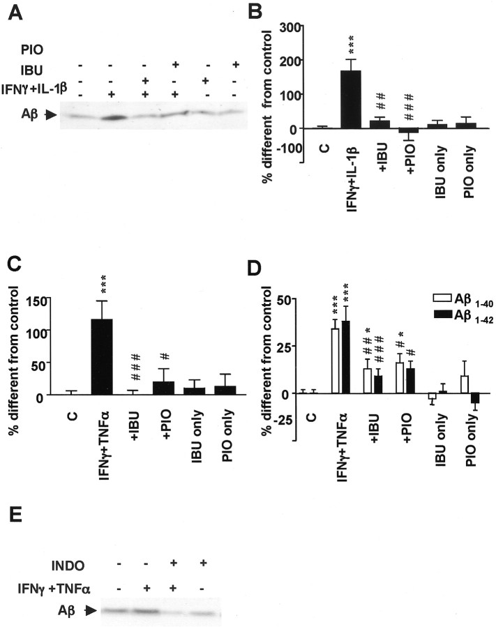
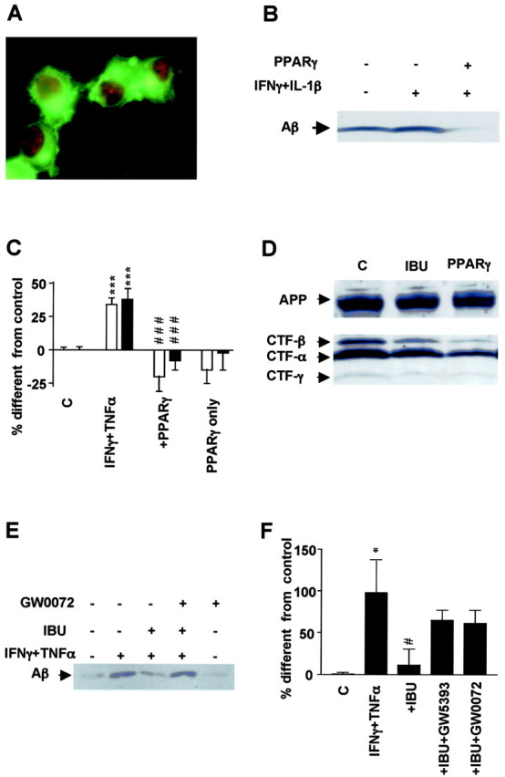
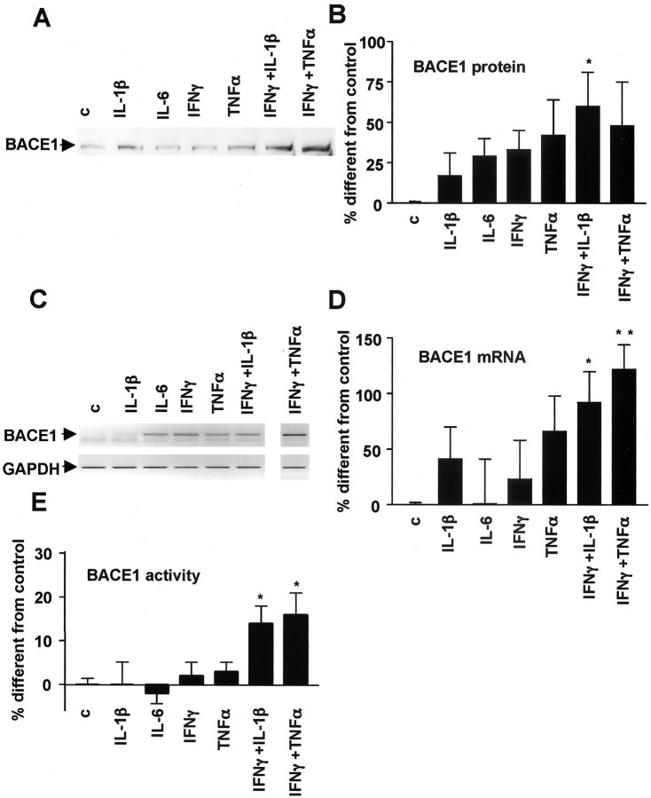
Long-term treatment with nonsteroidal anti-inflammatory drugs (NSAIDs) reduces the risk for Alzheimer's disease (AD). To determine the mechanisms by which inflammation affects AD and how NSAIDs protect against it, we stimulated neuroblastoma cells stably transfected with amyloid precursor protein (APP) with proinflammatory cytokines, which increased the secretion of amyloid-beta and APP ectodomain. Addition of ibuprofen, indomethacin, peroxisome proliferator-activated receptor-gamma (PPARgamma) agonists, or cotransfection with PPARgamma cDNA reversed this effect. The inhibitory action of ibuprofen and indomethacin was suppressed by PPARgamma antagonists. Finally, we observed that the mRNA levels, expression, and enzymatic activity of beta-secretase were increased by immunostimulation and normalized by NSAIDs. In conclusion, proinflammatory cytokines activate beta-secretase, and NSAIDs inhibit this effect through PPARgamma.


Figures

Figure 1.
Figure 1.
Proinflammatory cytokines affect sAPP secretion. A, Proinflammatory cytokines increase total sAPP (APPs) and α-sAPP (α-APPs) levels in N2a cells stably transfected with APPsw and immunostimulated with proinflammatory cytokines. B, Quantification of total sAPP andα-sAPP in N2a cells permanently transfected with APPsw by Western blotting experiments shows an increase after cytokine stimulation in six different experiments. C, Quantification of intracellular full-length APP in N2a cells permanently transfected with APPsw by Western blotting in nine experiments shows no effect after cytokine stimulation. Columns represent average ± SEM. *p ≤ 0.05; ANOVA, Tukey's post hoc test. c, Control.
Figure 2.
Figure 2.
Proinflammatory cytokines affect Aβ secretion. A, Increased Aβ generation in media obtained from N2a cells overexpressing APPsw by proinflammatory cytokines, shown by immunoprecipitation and Western blotting. B, Quantification of total Aβ by immunoprecipitation–Western blotting in five similar experiments as above. C, Analysis of Aβ1–40 and Aβ1–42 secretion in N2a APPsw cells by ELISA (n = 6). Columns represent average ± SEM. *p ≤ 0.05; **p ≤ 0.01; ***p ≤ 0.001; ANOVA, Tukey's post hoc test. c, Control.
Figure 3.
Figure 3.
Effect of Ibuprofen (IBU) and pioglitazone (PIO) on APP secretion. A, IBU and PIO decrease total sAPP (APPs) and α-sAPP (α-APPs) levels in N2a cells immunostimulated with proinflammatory cytokines but not under noninflammatory conditions. B, Quantification of sAPP by Western blot in six different experiments. C, Quantification of intracellular full-length APP by Western blotting shows no difference among treatments. Columns represent average ± SEM. Asterisks indicate significant differences between control and treatment. Number signs indicate significant differences between treatment with cytokines alone and with NSAIDs. **p ≤ 0.01; ***p ≤ 0.001; #p ≤ 0.05; ###p ≤ 0.001; ANOVA followed by a Tukey's post hoc test. C, Control.
Figure 4.
Figure 4.
Effect of ibuprofen (IBU) and pioglitazone (PIO) on Aβ secretion. A, Ibuprofen and pioglitazone decrease Aβ secretion in immunoprecipitation–Western blotting experiments in N2a cells for which APPsw was stimulated with IFNγ (1ng/ml)plus IL-1β (10 ng/ml) overnight and then incubated with IBU (10μm) or PIO (10μm) for 4 hr. B, Quantification of total Aβ in six to nine experiments of cells stimulated with the same conditions as above. C, Quantification of total Aβ in six to nine experiments of cells stimulated with IFNγ (1 ng/ml) plus TNFα (30 ng/ml). D, Analysis of Aβ1–40 and Aβ1–42 secretion in APPsw-transfected N2A cells by ELISA. E, Indomethacine (INDO) exerts the same effect as ibuprofen on Aβ generation. Columns represent average ± SEM. Asterisks indicate significant differences between control and treatment. Number signs indicate significant differences between treatment with cytokines alone and with NSAIDs. *p ≤ 0.05; ***p ≤ 0.001; #p ≤ 0.05; ##p ≤ 0.01; ###p ≤ 0.001; ANOVA followed by a Tukey's post hoc test. C, Control.
Figure 5.
Figure 5.
Effect of PPARγ on APP processing and Aβ secretion. A, Immunofluorescence detection of APP with 5313 antibody (green) and PPARγ with E-8 (red) in N2a cells overexpressing both proteins. B, Aβ detection by immunoprecipitation and Western blotting from media obtained from N2a cells overexpressing APPsw with or without transient transfection with PPARγ cDNA. PPARγ expression decreases generation of Aβ in cells stimulated overnight with the proinflammatory cytokines IFNγ (1 ng/ml) plus IL-1β (10 ng/ml). C, Analysis of Aβ1–40 and Aβ1–42 secretion by ELISA under the same conditions as above but in cells immunostimulated with IFNγ (1 ng/ml) plus TNFα (30 ng/ml). D, Detection of different CTFs of APP in N2a cells overexpressing APPsw and cotransfected with vector or PPARγ cDNA or incubated with ibuprofen (IBU). E, Total Aβ detection in N2A cells stimulated overnight with IFNγ plus TNFα and afterward with IBU (1μm) with or without PPARγ antagonist GW420072X (1μm) for 4 hr. F, Quantification of Aβ levels in eight experiments in which incubation with PPARγ antagonists GW0072X (1 μm) and GW5393X (1 μm) reversed the suppressive effect of ibuprofen (IBU) on APP processing. Columns represent average ± SEM. Asterisks indicate significant differences between control and treatment. Number signs represent differences between cytokines and NSAIDs. *p ≤ 0.05; ***p ≤ 0.001; #p ≤ 0.05; ###p ≤ 0.001; ANOVA followed by a Tukey's post hoc test. C, Control.
Figure 6.
Figure 6.
Effect of proinflammatory cytokines and NSAIDs on γ-secretase activity. A, No direct effect of NSAIDs onγ-secretase activity demonstrated by in vitro detection of CTF-γ from membrane preparations incubated for 1 hr with different concentrations of ibuprofen (IBU) or pioglitazone (PIO) was observed. B, Quantification of CTF-γ detected in vitro from three to five experiments. C, Aβ detection in N2A cells transfected with CT-99 mutant of APP shows no change with different treatments. D, Quantification of Aβ from four to five experiments from N2A cells transiently transfected with CT-99. E, Quantification of the ratio Aβ1–40/Aβ1–42from ELISA experiments performed in N2A transfected with APPsw cells shows an increase in this ratio after IBU treatment. Columns represent average ± SEM. Asterisks indicate significant differences. *p ≤ 0.05; ANOVA followed by a Tukey's post hoc test. C, Control.
Figure 7.
Figure 7.
Effect of proinflammatory cytokines on BACE1 expression, mRNA levels, and activity. A, Proinflammatory cytokines increase BACE1 expression in SK-N-SH cells by Western blotting experiments.B, Quantification of BACE1 expression in eight to nine experiments performed in N2A cells. C, Proinflammatory cytokines increase BACE1 steady-state mRNA levels shown in this semiquantitative RT-PCR representation from N2a cells treated with different proinflammatory cytokines. As a control, PCR was performed for GAPDH with the same samples. D, Quantification of BACE1 steady-state mRNA levels in six to nine experiments performed in N2A cells. E, Proinflammatory cytokines increase BACE1 activity in N2A cells. Columns represent average ± SEM. *p ≤ 0.05; **p < 0.01. c, Control.
Figure 8.
Figure 8.
Effect of ibuprofen (IBU) and pioglitazone (PIO) on BACE1 expression, mRNA levels, and activity. A, Increased BACE1 expression in SK-N-SH cells incubated with IFNγ (1 ng/ml) plus TNFα (30ng/ml) is reversed with IBU (10μm) in Western blotting experiments. B, Quantification of BACE1 expression in four to five experiments performed in N2A cells. Cells were incubated with IFNγ (1 ng/ml) plus TNFα (30 ng/ml) with or without IBU (10μm) or PIO(10 μm). C, Modulation of BACE1 steady-state mRNA levels by the same treatments is shown in semiquantitative RT-PCR representation of BACE1 mRNA from SK-N-SH cells. As a negative control, PCR was performed for β-actin with the same samples. D, PPARγ antagonists GW0072X (1 μm) reversed the effect of IBU and indomethacin (INDO) on BACE1 mRNA levels. E, Quantification of BACE1 steady-state mRNA levels in four to five experiments performed in N2A cells. F, BACE1 activity from N2A cells is affected by treatment with similar cytokines and NSAIDs. Asterisks indicate significant differences between control and treatment. Number signs represent differences between cytokines and NSAIDs. *p < 0.05. Columns represent average ± SEM; #p ≤ 0.05; ##p ≤ 0.01; ###p ≤ 0.001; ANOVA followed by a Tukey's post hoc test. C, Control.

References

    1. Aisen PS, Schafer KA, Grundman M, Pfeiffer E, Sano M, Davis KL, Farlow MR, Jin S, Thomas RG, Thal LJ ( 2003) Effects of rofecoxib or naproxen vs placebo on Alzheimer disease progression: a randomized controlled trial. JAMA 289: 2819–2826. - PubMed
    1. Akiyama H, Barger S, Barnum S, Bradt B, Bauer J, Cole GM, Cooper NR, Eikelenboom P, Emmerling M, Fiebich BL, Finch CE, Frautschy S, Griffin WS, Hampel H, Hull M, Landreth G, Lue L, Mrak R, Mackenzie IR, McGeer PL, et al. ( 2000) Inflammation and Alzheimer's disease. Neurobiol Aging 21: 383–421. - PMC - PubMed
    1. Alafuzoff I, Overmyer M, Helisalmi S, Soininen H ( 2000) Lower counts of astroglia and activated microglia in patients with Alzheimer's disease with regular use of non-steroidal anti-inflammatory drugs. J Alzheimers Dis 2: 37–46. - PubMed
    1. Amara FM, Junaid A, Clough RR, Liang B ( 1999) TGF-beta(1), regulation of alzheimer amyloid precursor protein mRNA expression in a normal human astrocyte cell line: mRNA stabilization. Brain Res Mol Brain Res 71: 42–49. - PubMed
    1. Avramovich Y, Amit T, Youdim MB ( 2002) Non-steroidal anti-inflammatory drugs stimulate secretion of non-amyloidogenic precursor protein. J Biol Chem 277: 31466–31473. - PubMed

Publication types

MeSH terms