. 2003 Oct 10;315(1):20-33.
doi: 10.1016/s0042-6822(03)00323-4.

Vacuolating encephalitis in mice infected by human coronavirus OC43

Affiliations

Vacuolating encephalitis in mice infected by human coronavirus OC43

Hélène Jacomy et al. Virology. .

Abstract

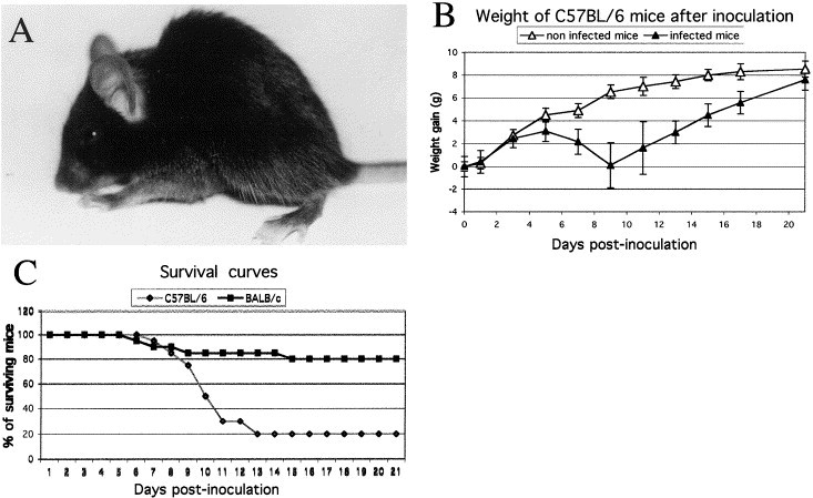
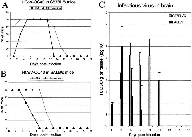
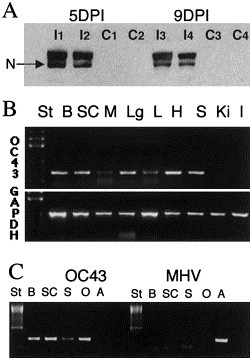
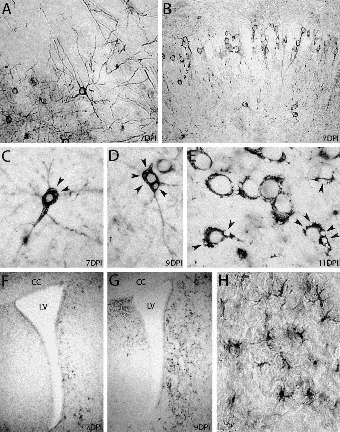
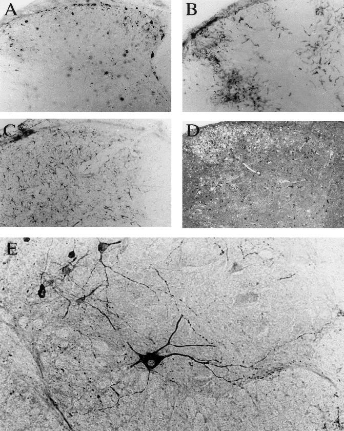
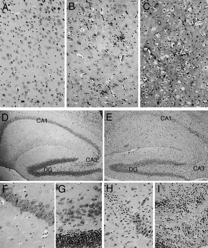
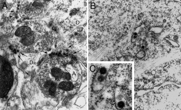
Involvement of viruses in human neurodegenerative diseases and the underlying pathologic mechanisms remain generally unclear. Human respiratory coronaviruses (HCoV) can infect neural cells, persist in human brain, and activate myelin-reactive T cells. As a means of understanding the human infection, we characterized in vivo the neurotropic and neuroinvasive properties of HCoV-OC43 through the development of an experimental animal model. Virus inoculation of 21-day postnatal C57BL/6 and BALB/c mice led to a generalized infection of the whole CNS, demonstrating HCoV-OC43 neuroinvasiveness and neurovirulence. This acute infection targeted neurons, which underwent vacuolation and degeneration while infected regions presented strong microglial reactivity and inflammatory reactions. Damage to the CNS was not immunologically mediated and microglial reactivity was instead a consequence of direct virus-mediated neuronal injury. Although this acute encephalitis appears generally similar to that induced by murine coronaviruses, an important difference rests in the prominent spongiform-like degeneration that could trigger neuropathology in surviving animals.


Figures

Fig. 1
Fig. 1
Characterization of HCoV-OC43 infection. (A) Typical posture of an infected mouse at an advanced stage of disease when the animal presented highly pronounced humped back with restrained mobility (9 DPN). (B) C57BL/6 mice were weighed every 2 days after infection to estimate weight variations. HCoV-OC43-infected mice gained weight normally during the first 5 days after infection, after which they all lost weight during the acute phase of the disease. The more affected mice lost more weight more rapidly than less affected mice and died during this period. After 9 days postinfection, mice which survived gained weight to reach the weight of control animals around 21 days postinfection. (C) Survival curves of mice after HCoV-OC43 infection. BALB/c mice received a higher dose than the C57BL/6 mice, 10,000 TCID50 versus 10 TCID50. However, C57BL/6 were less resistant, with 80 versus 20% of death after infection.
Fig. 2
Fig. 2
HCoV-OC43 infectious virus and RNA in the CNS of 21 DPN mice. (A) 100% of brains from C57BL/6 mice inoculated ic with 10 TCID50 of HCoV- OC43 were positive for viral RNA between 3 and 11 days postinfection. Only 25% of the surviving mice were found positive after 15 days postinfection and RNA was not found thereafter. Infectious virus appeared later and disappeared before elimination of viral RNA. Between 5 and 11 days postinfection, 100% of brains contained infectious virus. (B) Detection of HCoV-OC43 RNA in the brain of BALB/c mice inoculated ic with 105 TCID50 of HCoV-OC43 revealed that 100% of these mice were positive until 9 days postinfection. Infectious virus was detectable in all mice only during the first 3 days postinfection and gradually fewer mice were found positive. (C) Histogram representing the amount of infectious virus detected in five brains from the two strains of mice at different intervals postinfection. The limit of the detection assay was 100.5 TCID50.
Fig. 3
Fig. 3
Detection of proteins and RNA in brains of C57BL/6 mice inoculated ic with HCoV-OC43 at 21 DPN. (A) Western blot analysis at 5 and 9 days postinfection, revealing the presence of the HCoV-OC43 N proteins in inoculated mice (I1, I2, I3, and I4 mice) and its absence in control mice (C1, C2, C3, and C4). (B) RT-PCR analysis revealed at 13 days postinfection that virus had spread from the CNS (B: brain; SC: spinal cord) to other peripheric organs, such as heart (H), lungs (Lg), spleen (S), and to a lesser extent, the liver (L) and muscles (M). HCoV-OC43 virus was never found in kidneys (Ki) nor in intestine (I). The quality of RNA extraction was estimated using GAPDH RNA detection. (C) RT-PCR analysis demonstrating that HCoV-OC43 and not an enzootic MHV strain was detected. HCoV-OC43 primers recognized HCoV-OC43 mRNA in B, SC, or S tissues of an infected mouse as well as in control RNA (O: HCoV-OC43-infected cells) but not MHV-A59 mRNA (A: MHV-infected cells). On the other hand, on the same sample, MHV primers recognized only MHV-A59 mRNA but not HCoV-OC43 mRNA.
Fig. 4
Fig. 4
Immunocytochemical staining of infected (21 DPN) C57BL/6 mice brains. HCoV-OC43-infected neurons stained in black in the cerebral cortex (A) and in the CA1 layer of the hippocampus (B), 1 week after virus inoculation. In C, D, and E, HCoV-OC43-positive neurons exhibited empty vacuoles (arrowheads) in the cytoplasm at 7 days postinfection, which increased at 9 and 11 days postinfection. Panels F, G, and H show reactive microgliosis in the brain. At 7 days postinfection (F), Mac-2-stained cells started to appear along the lateral ventricle (LV) and became more numerous at 9 days postinfection (G). H shows a magnification of microglial cells observed in G (CC: corpus callosus). A, B, H: original magnification: ×200; F,G: magnification: ×40; C, D, E: ×400.
Fig. 5
Fig. 5
Immunocytochemical staining of the spinal cord of a C57BL/6 mouse (21 DPN) 1 week after HCoV-OC43 infection. (A, B, and C) Evidence of inflammatory reaction on three consecutive sections of the cervical dorsal horn. (A): Antiviral MAb. (B) Mac-2 microglia/macrophages cell marker. (C): GFAP astrocytic cell marker. (D): spongiosis aspect of gray matter revealed by toluidine blue staining. (E): Infection of motor neurons by HCoV-OC43 in the ventral horn of the lumbar spinal cord. Magnification: ×100 before enlargement.
Fig. 6
Fig. 6
Cytotoxic effect of HCoV-OC43 infection in the CNS of C57BL/6 mice (21 DPN) at 11 days postinfection (hematoxylin and eosin staining) (A, B, and C) Overview of the thalamus (original magnification: ×200). (A) Control mice showing no degenerative changes. B (less affected animal) and C (more affected animal) illustrate infiltrating cells and spongiform-like lesions (characterized by vacuolation in neuronal cell bodies) and degenerative neurons with small densely stained nuclei or eosinophilic cytoplasm. (D) Normal hippocampus from control mice, with magnification of CA1 (F) and CA3 (G) hippocampus layers. (E) After infection, degenerative changes appeared mostly in CA1 (H) and CA3 (I) hippocampal layers; degenerated neurons exhibited picnotic nuclei and vacuolated cells were present in CA3 hippocampal layers (DG: Dentate Gyrus). (D and E) Magnification: ×40; F, G, H, and I: original magnification: ×200.
Fig. 7
Fig. 7
Detection of HCoV-OC43 infection at the electron microscopic level, in the spinal cord of C57BL/6 mice infected 7 days previously. (A) Numerous viral particles (arrows) were seen between the axons (A) and the dendrites (D). (B) Virus particles found in the endoplasmic reticulum of the cytoplasm (original magnification: ×20,000). (C) Magnification of B to show virions. Viral spikes around virions are typical of coronaviruses. No retroviral particles were observed (original magnification: ×40,000).
Fig. 8
Fig. 8
Survival curves of infected mice treated with cyclosporin A (CsA). Group of 10 C57BL/6 mice treated with cyclosporin A at 20 mg/kg/day (OC43/20 mgCsA) and at 10 mg/kg/day (OC43/10 mgCsA) became more susceptible to HCoV-OC43 infection, with 100 and 90% of death after infection versus 80% in non-CsA-treated animal. Infected C57BL/6 mice treated with oil alone (OC43/oil) presented similar survival curves as previously reported, with 80% of death after infection. Noninfected HCoV-OC43 mice treated with CsA at 20 mg/kg/day (Control/20 mgCsA) illustrated that CsA was not toxic under these conditions.

References

    1. Agapitos E., Pavlopoulos P.M., Patsouris E., Davaris P. Subacute necrotizing encephalopathy (Leigh's disease): a clinicopathologic study of ten cases. Gen. Diag. Pathol. 1997;142:341–355. - PubMed
    1. Allen I.V., McQuaid S., McMahon J., Kirk J., McConnell R. The significance of measles virus antigen and genome distribution in the CNS in SSPE for mechanisms of viral spread and demyelination. J. Neuropathol. Exp. Neurol. 1996;55:471–480. - PubMed
    1. Arbour N., Côté G., Lachance C., Tardieu M., Cashman N.R., Talbot P.J. Acute and persistent infection of human neural cell lines by human coronavirus OC43. J. Virol. 1999;73:3338–3350. - PMC - PubMed
    1. Arbour N., Day R., Newcombe J., Talbot P.J. Neuroinvasion by human respiratory coronaviruses and association with multiple sclerosis. J. Virol. 2000;74:8913–8921. - PMC - PubMed
    1. Arbour N., Ekandé S., Côté G., Lachance C., Chagnon F., Tardieu M., Cashman N.R., Talbot P.J. Persistent infection of human oligodendrocytic and neuroglial cell lines by human coronavirus 229E. J. Virol. 1999;73:3326–3337. - PMC - PubMed

Publication types

MeSH terms