. 2003 Nov 3;22(21):5723-33.
doi: 10.1093/emboj/cdg560.

E-cadherin controls adherens junctions in the epidermis and the renewal of hair follicles

Affiliations

E-cadherin controls adherens junctions in the epidermis and the renewal of hair follicles

Peter Young et al. EMBO J. .

Abstract

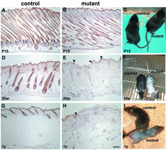
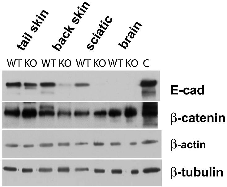
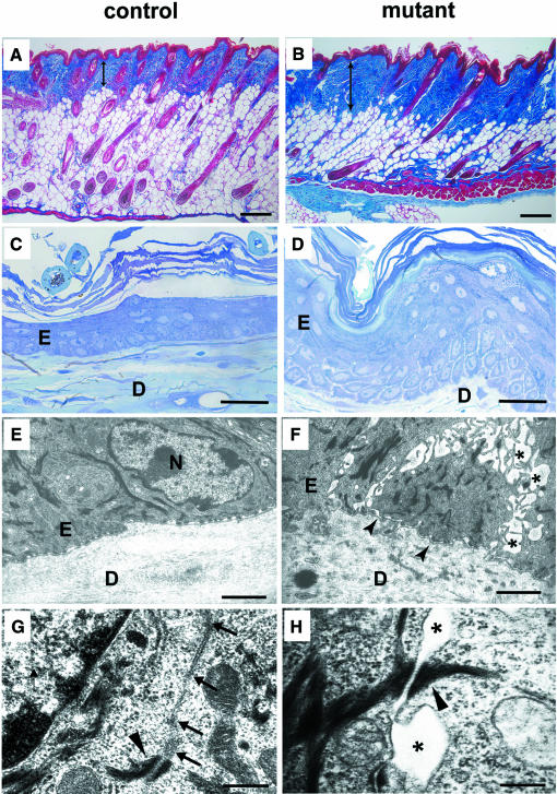
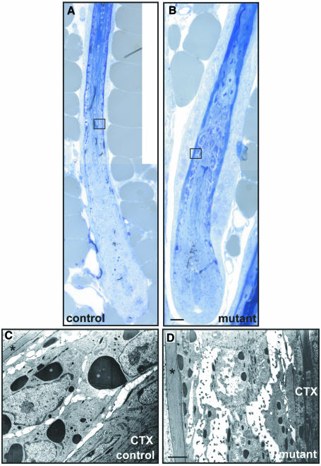
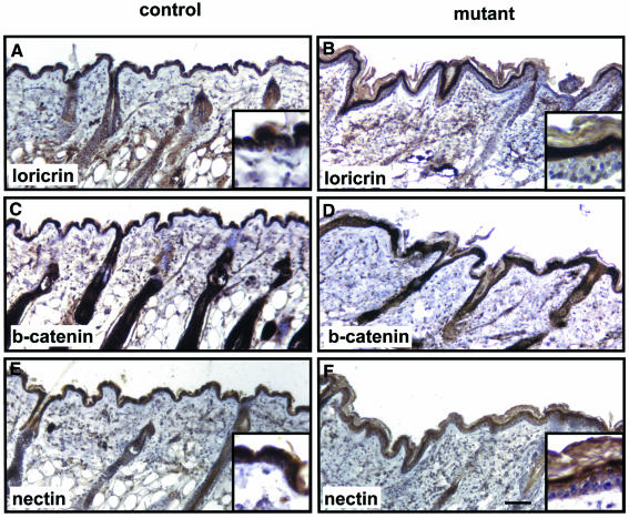
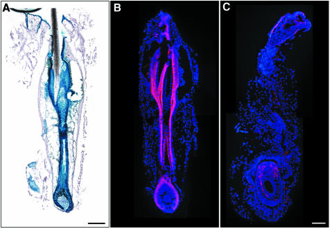
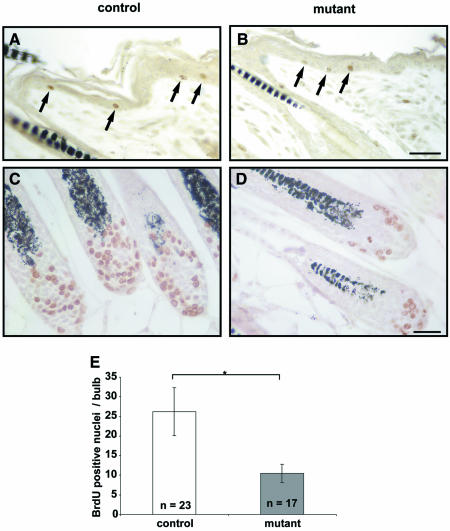
E-cadherin is thought to mediate intercellular adhesion in the mammalian epidermis and in hair follicles as the adhesive component of adherens junctions. We have tested this role of E-cadherin directly by conditional gene ablation in the mouse. We show that postnatal loss of E-cadherin in keratinocytes leads to a loss of adherens junctions and altered epidermal differentiation without accompanying signs of inflammation. Overall tissue integrity and desmosomal structures were maintained, but skin hair follicles were progressively lost. Tumors were not observed and beta-catenin levels were not strongly altered in the mutant skin. We conclude that E-cadherin is required for maintaining the adhesive properties of adherens junctions in keratinocytes and proper skin differentiation. Furthermore, continuous hair follicle cycling is dependent on E-cadherin.


Figures

None
Fig. 1. Ablation of E-cadherin in back skin epidermis and in hair follicles of mutant mice. Epidermis of back skin of P15 control (A) and mutant (B) mice shows a reduction of E-cadherin immunoreactivity in the upper parts of the hair follicles of mutant mice, while no difference can be detected in the epidermis. At the age of 20 weeks (20w), mutant mice show a massive reduction (78%) of the E-cadherin immuno-positive area within the epidermis, and the hair follicles have lost E-cadherin completely (E). In control animals, E-cadherin was detected throughout the epidermis and in all hair follicles (D). One-year-old (1y) mutant mice displayed a further increase in E-cadherin-negative areas (92%) (H). In control animals, E-cadherin staining was localized in the epidermis and hair follicles (G). Phenotypically, mutant mice were indistinguishable from control mice at P15 (C). Hair loss was already close to maximal at 20 weeks (F) and progressed slightly (I). The age of the animals is indicated in the lower left corner of each figure. Arrowheads indicate remaining E-cadherin-positive epidermal cells in mutant animals. Scale bar in (H) (also for A, B, D, E and G): 100 µm.
None
Fig. 2. Western blot analysis of E-cadherin expression in different tissues from 20-week-old control (WT) and mutant (KO) mice. Protein was extracted from back skin, tail skin, sciatic nerve and whole brain from control and mutant mice. E-cadherin levels were strongly reduced in back skin and sciatic nerve of mutant animals. Tail skin showed comparable amounts of E-cadherin.
None
Fig. 3. Monitoring of recombination in Krox20Cre/+; R26R reporter mice by X-gal staining for β-galactosidase (β-gal) activity in the back skin at different postnatal developmental stages. At P1, recombination had occurred within only very small epidermal patches located near the hair follicle (A). In later stages (P10 and P18), the upper parts of the hair follicles show β-gal-positive cells, and β-gal-positive patches within the epidermis increased (B and C). At the age of 20 weeks (20w), all hair follicles and most parts of the epidermis show β-gal-positive cells indicating recombination (D). Scale bar in (D) (also for A–C): 200 µm.
None
Fig. 4. Morphological changes resulting from ablation of the E-cadherin gene in the skin and hair follicles in adult 20-week-old mice. Masson trichrome staining revealed increased collagen deposition (indicated by the double arrowed line), and a reduction of hair follicles in mutant animals (B) in comparison with control animals (A). In contrast to control epidermis (C), mutant animals displayed strong epidermal acanthosis and mild parakeratosis (D). A widening of intercellular spaces was evident in the basal cell layer and the layers just above. On the ultrastructural level, widening of intracellular spaces was seen in the basal epidermal cell layer in back skin of mutant mice (asterisks) (F). Basally localized hemidesmosomes were detectable in control (E) as in mutant animals (F), but the basement membrane was undulated in mutant animals, as indicated by arrowheads (F). At higher magnification, loss of adherens junctions was seen in mutant animals indicated by asterisks (H), while control mice showed normal adherens junctions (G; arrows). Desmosomes in the epidermis of mutant animals were preserved (H; arrowhead). E = epidermis, D = dermal compartment, N = nucleus. Scale bars in (A–D), 10 µm; (E and F), 2 µm; (G and H), 200 nm.
None
Fig. 5. Loss of intercellular tightness between cells of the Huxley layer of the IRS in back skin. Analysis of the remaining hair follicles in the back skin of 20-week-old mutant mice revealed a loss of intercellular tightness in the Huxley layer of the IRS (B and D). As in adult control animals, the Henle layer (indicated by asterisks) was not disturbed and the cortex of the hair was normal (A and B). Trichohyalin granula were massively reduced in mutant mice. Note the supernumery cells in the ORS of mutant mice (B). CTX = cortex of the hair shaft. Scale bar in (B), 50 µm (for A and B); (D) 2 µm (for C and D).
None
Fig. 6. Keratinocyte differentiation in adult E-cadherin mutant mice. Sections from back skin of control (A, C and E) and mutant (B, D and F) mice were stained with antibodies against K6 (green), E-cadherin (E-cad; red), desmoplakin (green), K14 (red) and K10 (green) as indicated. K6 expression was restricted to the hair follicle in control mice (A), whereas in mutant mice it was also ectopically found in the interfollicular epidermis (B). Desmoplakin was localized in all epidermal layers in control (C) and in mutant mice (D). K14 was found in the basal layer in back skin of control mice (E) and mutants (F). The extended expression of K14 in the mutants reflects mainly the enlargement (hypertrophy) of the basal cell layer. Note the acanthosis of the epidermis in all sections from mutant mice. Scale bar in (B), 100 µm (for A and B); (F), 50 µm (for C–F).
None
Fig. 7. Epidermal expression of loricrin, β-catenin and nectin 1α in back skin of mutant E-cadherin-deficient mice. The epidermis of back skin from 20-week-old mutant mice showed loricrin-negative supernumery cells above the basal keratinocyte layer (A and B). β-catenin was expressed throughout the epidermis in control and mutant animals (C and D). Additional nectin 1α-negative (or low expressing) cells were found in the suprabasal region of mutant animals (F) compared with control animals (E). Scale bar in (F), 100 µm (also for A–E).
None
Fig. 8. Abnormal morphology in micro-dissected whisker follicles of adult (20-week-old) mutant mice. Recombination was detectable throughout the entire whisker follicle in Krox20Cre/+; R26R β-gal-reporter mice (A). E-cadherin expression was observed in control animals in the outer root sheath and parts of the inner root sheath (pink; B). In mutant animals, E-cadherin was almost totally lost and only a few E-cadherin-positive cells remained in the hair bulb (pink, C). Sections in (B) and (C) were counterstained with DAPI to visualize nuclei. Scale bar in (A and B), 50 µm (for A–C).
None
Fig. 9. Reduced proliferation in hair bulbs of 15-day-old E-cadherin-deficient mice. Some BrdU-labeled proliferating cells were observed in the epidermis of control (A) and mutant animals (B). In the hair bulbs, the number of proliferating cells was significantly reduced in mutant animals (D) compared with control animals (C). (E) Quantitative analysis of three independent experiments. Cells of medially cut bulbs were counted. The asterisk indicates statistical significance (P < 0.005). Scale bar in (B), 100 µm (for A and B), (D), 20 µm (for C and D).

References

    1. Aberle H., Schwartz,H. and Kemler,R. (1996) Cadherin–catenin complex: protein interactions and their implications for cadherin function. J. Cell. Biochem., 61, 514–523. - PubMed
    1. Boussadia O., Kutsch,S., Hierholzer,A., Delmas,V. and Kemler,R. (2002) E-cadherin is a survival factor for the lactating mouse mammary gland. Mech. Dev., 115, 53–62. - PubMed
    1. Braga V. (2000) Epithelial cell shape: cadherins and small GTPases. Exp. Cell Res., 261, 83–90. - PubMed
    1. Brakebusch C. et al. (2000) Skin and hair follicle integrity is crucially dependent on β1 integrin expression on keratinocytes. EMBO J., 19, 3990–4003. - PMC - PubMed
    1. Burge S.M. (1992) Hailey–Hailey disease: the clinical features, response to treatment and prognosis. Br. J. Dermatol., 126, 275–282. - PubMed

Publication types

MeSH terms