. 2003 Nov 3;22(21):5734-45.
doi: 10.1093/emboj/cdg545.

PP1 control of M phase entry exerted through 14-3-3-regulated Cdc25 dephosphorylation

Affiliations

PP1 control of M phase entry exerted through 14-3-3-regulated Cdc25 dephosphorylation

Seth S Margolis et al. EMBO J. .

Abstract

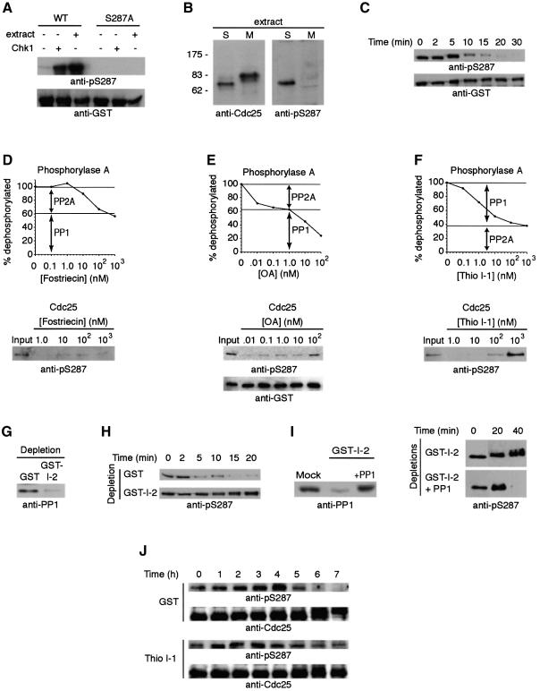
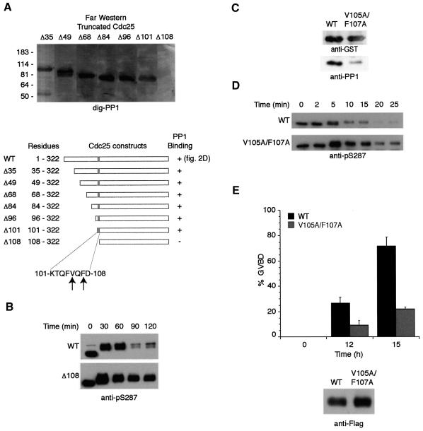
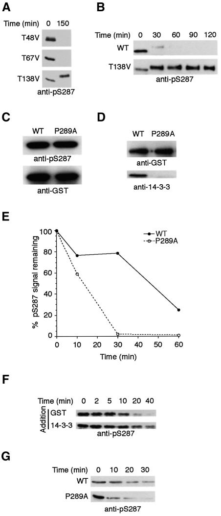
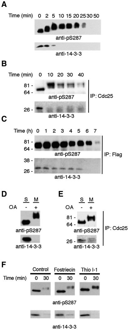
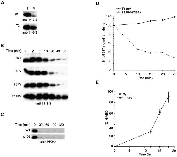
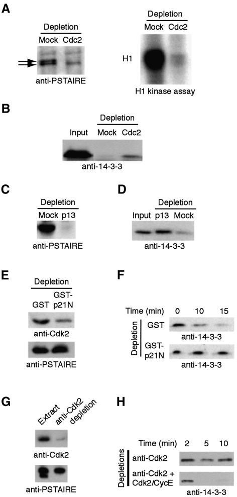
It has been known for over a decade that inhibition of protein phosphatase 1 (PP1) activity prevents entry into M phase, but the relevant substrate has not been identified. We report here that PP1 is required for dephosphorylation of the Cdc2-directed phosphatase Cdc25 at Ser287 (of Xenopus Cdc25; Ser216 of human Cdc25C), a site that suppresses Cdc25 during interphase. Moreover, PP1 recognizes Cdc25 directly by interacting with a PP1-binding motif in the Cdc25 N-terminus. We have also found that 14-3-3 binding to phospho-Ser287 protects Cdc25 from premature dephosphorylation. Upon entry into M phase, 14-3-3 removal from Cdc25 precedes Ser287 dephosphorylation, suggesting the existence of a phosphatase- independent pathway for 14-3-3 removal from Cdc25. We show here that this dissociation of 14-3-3 from Cdc25 requires the activity of the cyclin-dependent kinase Cdk2, providing a molecular explanation for the previously reported requirement for Cdk2 in promoting mitotic entry. Collectively, our data clarify several steps important for Cdc25 activation and provide new insight into the role of PP1 in Cdc2 activation and mitotic entry.


Figures

None
Fig. 1. S287 is dephosphorylated by PP1. (A) Full-length GST–Cdc25 and GST–Cdc25 S287A proteins were incubated with Xenopus interphase egg extract at 4°C or Chk1 + ATP at 30°C for 1.5 h. Samples were collected, washed and resolved by SDS–PAGE for immunoblotting with either anti-pSer287 or anti-GST antibodies. (B) Total interphase or mitotic extracts were resolved by SDS–PAGE and immunoblotted with the indicated antisera. (C) Following GST–Cdc25 incubation in interphase egg extract, non-degradable cyclin was added to drive the extract into mitosis. GST–Cdc25 was retrieved from the extract at the indicated times using glutathione–Sepharose beads. Beads were washed extensively with egg lysis buffer (ELB) and boiled in sample buffer for SDS–PAGE and immunoblotting with the indicated antibodies. (DF) Top: radiolabeled phosphorylase A was incubated in mitotic egg extract in the presence of increasing concentrations of fostriecin (D), okadaic acid (E) or thiophosphorylated I-1 (F). The percentage of phosphorylase A which was dephosphorylated is shown. Inhibition of PP2A by fostriecin allows 60% dephosphorylation of phosphorylase A by PP1, while PP1 inhibition by thio-I-1 allows 40% dephosphorylation by PP2A. Similar ratios of PP1 and PP2A-dependent dephosphorylation are observed with okadaic acid titration. Lower panels: GST–Cdc25 (amino acids 1–322) was pre-phosphorylated with Chk1 in kinase buffer with ATP at 30°C for 1.5 h, washed and then incubated with mitotic egg extract and increasing amounts of fostriecin, okadaic acid or thio-I-1, as indicated. Samples were processed for anti-pS287 or anti-GST immunoblotting. (G) PP1 was depleted from mitotic egg extracts using GST–I-2–Sepharose or mock depleted using GST–Sepharose. Depleted extracts were resolved by SDS–PAGE and immunoblotted with anti-PP1 sera. (H) Full-length GST–Cdc25 was incubated in interphase extract, retrieved on glutathione–beads, washed with ELB and placed into either GST or GST–I-2-depleted extracts. Samples were collected at the indicated times, washed, boiled in sample buffer for SDS–PAGE and then immunoblotted with anti-pS287 antibodies. (I) Left: extracts depleted as in (G) were immunoblotted with anti-PP1 before and after re-addition of pure PP1. Right: samples were processed as in (H) in the presence and absence of reconstituted pure PP1. (J) Stage VI oocytes were injected with either GST or thio-I-1 followed by progesterone treatment. Twenty oocytes were taken at the indicated times and frozen in liquid nitrogen. Samples were lysed, and endogenous Cdc25 was detected by anti-pS287 and anti-Cdc25 immunoblotting.
None
Fig. 2. PP1 binds directly to Cdc25. (A) GST or GST–Cdc25 linked to glutathione–Sepharose was dipped in mitotic egg extracts, retrieved by centrifugation, washed and immunoblotted with anti-PP1 antibody. Coomassie-stained gel of pull-downs showed equal loading of GST and GST–Cdc25. The same protein preparations were used in (E), below. (B) Immunoprecipitates formed from mitotic egg extracts using either anti-Cdc25 or pre-immune sera were resolved by SDS–PAGE and immunoblotted with either anti-Cdc25 or anti-PP1 antibodies. The small arrowhead denotes the IgG heavy chain. (C) Mitotic extract samples were immunoprecipitated with anti-PP1 monoclonal or control IgG and blotted with either anti-Cdc25 or anti-PP1 antibodies. The small arrowhead denotes the IgG light chain. (D) Sepharose beads linked to 0.1 µg of GST–Cdc25 (amino acids 1–322) or GST were incubated in 100 µl egg extracts supplemented with non-degradable cyclin. After 30 min, samples were retrieved by centrifugation, washed, resolved by SDS–PAGE, transferred to PVDF membrane and probed with dig-PP1 (as in Terry-Lorenzo et al., 2000). (E) GST or GST–Cdc25 linked to glutathione–Sepharose was incubated with purified PP1 in vitro. Samples were collected by centrifugation, washed, and prepared for anti-PP1 immunoblotting.
None
Fig. 3. Cdc25 contains a consensus PP1-binding site important for efficient S287 dephosphorylation. (A) An N-terminal truncation series of GST–Cdc25 proteins was constructed and probed for PP1 binding by far western blotting with dig-PP1. (B) The GST–Δ108 mutant and WT GST–Cdc25 proteins were incubated in Xenopus egg extracts followed by addition of non-degradable cyclin. Proteins were retrieved from extracts with glutathione–Sepharose and immunoblotted with anti-pS287 antibody. (C) WT and V105A/F107A Cdc25 proteins were assayed for their ability to bind PP1 from egg extract as in Figure 2A. Samples were also blotted with anti-GST to show equal loading of baits. (D) GST–WT Cdc25 and V105A/F107A proteins were treated as in (B). (E) mRNA encoding Flag-tagged WT or V105A/F107A Cdc25 was injected into oocytes treated with the nuclear export inhibitor leptomycin B (200 nM), which enhances Cdc25 potency. Oocytes were monitored for GVBD over time. At 15 h incubation, 30 oocytes were collected, lysed, mixed with sample buffer, and resolved by SDS–PAGE for immunoblotting with anti-Flag antibodies.
None
Fig. 4. S287 dephosphorylation is regulated by 14-3-3 binding. (A) T138V, T48V and T67V GST–Cdc25 (1–322) proteins were incubated in interphase extract supplemented with non-degradable cyclin. Cdc25 proteins were retrieved on glutathione–Sepharose at the indicated times and immunoblotted with anti-pS287 antibody. (B) Full-length WT or T138V mutant GST–Cdc25 proteins were tested as in (A). (C) GST–P289A and WT Cdc25 proteins were incubated with Chk1 in kinase buffer and ATP to assess comparative abilities of the P289A and WT Cdc25 proteins to be phosphorylated at S287. Samples were immunoblotted with anti-pS287 or anti-GST antibodies. (D) GST–P289A and WT Cdc25 proteins were incubated in egg extract for 1 h at 4°C. Samples were retrieved on glutathione–Sepharose, washed with ELB plus 0.1% Triton X-100 and 300 mM NaCl, and resolved by SDS–PAGE to blot for 14-3-3 or GST. (E) GST–P289A and WT Cdc25 proteins were incubated in interphase extract followed by addition of non-degradable cyclin. Cdc25 proteins were retrieved on glutathione–Sepharose at the indicated times and immunoblotted with anti-pSer287 antibodies. Signals quantitated by densitometry are represented as percentage pS287 signal remaining. (F) GST–Cdc25 (amino acids 1–322) was pre-phosphorylated using Chk1 and then incubated with PP1 in vitro in the presence of GST or 14-3-3 at 37°C. Samples were taken at the indicated times, washed, and immunoblotted with anti-pS287. (G) Samples were processed as in (F), but P289A and WT proteins were compared in the presence of recombinant 14-3-3.
None
Fig. 5. 14-3-3 removal occurs prior to and independently of S287 dephosphorylation. (A) GST–Cdc25 phosphorylated by Chk1 and bound to 14-3-3 was added to M phase egg extract. Cdc25 was retrieved from the extract at the times shown, resolved by SDS–PAGE, and immunoblotted with either anti-14-3-3 antibody or anti-pSer 287 antibody. (B) Endogenous Cdc25 was immunoprecipitated from interphase extract and transferred into mitotic extracts. At the times indicated, the precipitates were then re-collected by centrifugation and processed for immunoblotting with either anti-pS287 or anti-14-3-3 antibody. (C) Stage VI oocytes were injected with mRNA encoding Flag-tagged Cdc25 and treated with progesterone. At the times indicated, 30 oocytes were collected, lysed, immunoprecipitated with anti-Flag, resolved by SDS–PAGE, and immunoblotted with anti-pS287 and anti-14-3-3 antibodies. (D) Cdc25 phosphorylated on S287 and loaded with 14-3-3 was transferred into M extract in the presence or absence of 5 µM okadaic acid. Samples were removed and immunoblotted with either 14-3-3 antibody or anti-pS287. Note that 14-3-3 was removed despite the maintenance of S287 phosphorylation. (E) Endogenous Cdc25 immunoprecipitated from interphase extract was transferred into mitotic extract supplemented with 5 µM okadaic acid and processed as in (D). (F) Samples were processed as in (D), but in the presence of fostriecin or thio I-1.
None
Fig. 6. T138 phosphorylation is required for 14-3-3 removal from Cdc25. (A) WT GST–Cdc25 or GST–Cdc25 in which Thr48, Thr67 and Thr138 had all been changed to valine were incubated in interphase extract to acquire S287 phosphorylation and bind 14-3-3. These samples were retrieved on glutathione–Sepharose and transferred to either interphase or mitotic egg extract. Samples were then re-precipitated and processed for anti-14-3-3 immunoblotting. (B) Individual mutant Cdc25 proteins were incubated in interphase extract, captured on glutathione–Sepharose and transferred to mitotic extract. Beads containing Cdc25 were pelleted at the times indicated, washed, and processed for anti-14-3-3 immunoblotting. (C) The samples shown in Figure 3B were immunoblotted with anti-14-3-3 antibodies. (D) The T138V and T138V/P289A double mutant Cdc25 proteins were incubated in interphase extract followed by addition of non-degradable cyclin. Cdc25 proteins were retrieved on glutathione–Sepharose at the indicated times and immunoblotted with anti-pS287 antibodies. Signals were quantitated by densitometry and are represented as percentage pS287 signal remaining. (E) Oocytes were injected with mRNA encoding WT or T138V Flag-tagged Cdc25 in the presence of 200 nM leptomycin B. Samples were monitored for GVBD over time.
None
Fig. 7. Cdk2 participates in 14-3-3 removal from Cdc25. (A) Mitotic extracts were immunodepleted using Sepharose linked to Xenopus Cdc2 antibodies and immunoblotted with anti-PSTAIRE monoclonal antibody [able to recognize both Cdc2 (upper band) and Cdk2 (lower band)]. Histone H1 kinase assays were performed on mock-depleted and Cdc2-depleted samples. (B) GST–Cdc25 bound to 14-3-3 was added to the anti-Cdc2 or mock-depleted extracts, retrieved on glutathione–Sepharose, washed, resolved by SDS–PAGE and immunoblotted with anti-14-3-3 antibody. (C) Mitotic egg extract was depleted on p13 Sepharose or mock depleted. Depleted extracts resolved by SDS–PAGE were immunoblotted with anti-PSTAIRE. (D) The extracts from (C) were processed for Cdc25–14-3-3 interactions as in (B). (E) GST linked to a N-terminal fragment of p21 protein or GST alone was bound to glutathione–Sepharose and used as a resin to deplete mitotic extracts. Depleted samples were immunoblotted with Cdk2 antibody. (F) Samples depleted as in (E) were supplemented with GST–Cdc25 bound to 14-3-3. The Cdc25 protein was retrieved from the extracts at the indicated times, washed, resolved by SDS–PAGE and immunoblotted with anti-14-3-3 antibody. (G) Mitotic extracts were immunodepleted using anti-Cdk2 antibodies linked to protein A–Sepharose. Depleted extracts were immunoblotted with anti-Cdk2 or anti-PSTAIRE (note Cdk2, but not Cdc2, is removed). (H) Cdk2-depleted samples from (G) in the presence or absence of reconstituted Cdk2–cyclin E were assayed for 14-3-3 release as in (F).

References

    1. Bulavin D.V. et al. (2003) Dual phosphorylation controls Cdc25 phosphatases and mitotic entry. Nat. Cell Biol., 5, 545–551. - PubMed
    1. Cohen P.T. (2002) Protein phosphatase 1-targeted in many directions. J. Cell Sci., 115, 241–256. - PubMed
    1. Coleman T.R. and Dunphy,W.G. (1994) Cdc2 regulatory factors. Curr. Opin. Cell Biol., 6, 877–882. - PubMed
    1. Connor J.H., Quan,H., Oliver,C. and Shenolikar,S. (1998) Inhibitor-1, a regulator of protein phosphatase 1 function. Methods Mol. Biol., 93, 41–58. - PubMed
    1. Duckworth B.C., Weaver,J.S. and Ruderman,J.V. (2002) G2 arrest in Xenopus oocytes depends on phosphorylation of cdc25 by protein kinase A. Proc. Natl Acad. Sci. USA, 99, 16794–16799. - PMC - PubMed

Publication types

MeSH terms