. 2003 Nov 3;22(21):5769-79.
doi: 10.1093/emboj/cdg548.

scribble mutants cooperate with oncogenic Ras or Notch to cause neoplastic overgrowth in Drosophila

Affiliations

scribble mutants cooperate with oncogenic Ras or Notch to cause neoplastic overgrowth in Drosophila

Anthony M Brumby et al. EMBO J. .

Abstract

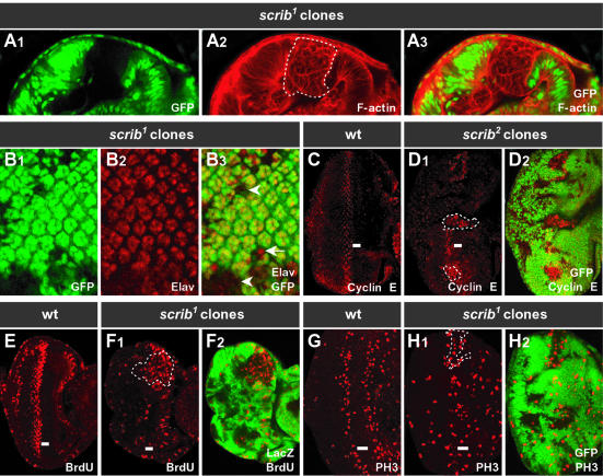
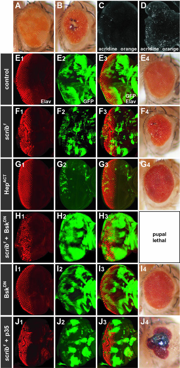
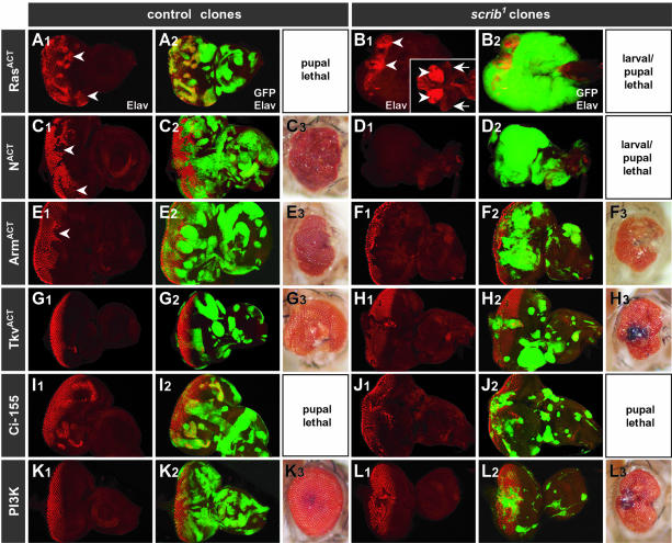
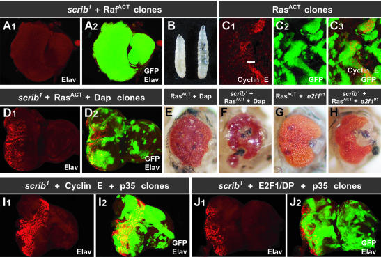
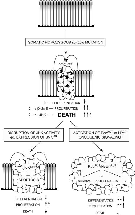
Cancer is a multistep process involving cooperation between oncogenic or tumor suppressor mutations and interactions between the tumor and surrounding normal tissue. Here we present the first description of cooperative tumorigenesis in Drosophila, by using a system that mimics the development of tumors in mammals. We have used the MARCM system to generate mutant clones of the apical-basal cell polarity tumor suppressor gene, scribble, in the context of normal tissue. We show that scribble mutant clones in the eye disc exhibit ectopic expression of cyclin E and ectopic cell cycles, but do not overgrow due to increased cell death mediated by the JNK pathway and the surrounding wild-type tissue. In contrast, when oncogenic Ras or Notch is expressed within the scribble mutant clones, cell death is prevented and neoplastic tumors develop. This demonstrates, for the first time in Drosophila, that activated alleles of Ras and Notch can act as cooperating oncogenes in the development of epithelial tumors, and highlights the importance of epithelial polarity regulators in restraining oncogenes and preventing tumor formation.


Figures

None
Fig. 1. Somatic eye clones of scrib mutant tissue become multilayered and overproliferate. (A) Cross-section and (B–H) planar-sections, anterior to the right, through the monolayered epithelium of third instar larval eye discs. ey-FLP scrib1 or scrib2 mutant clones (sometimes outlined by dashed lines) are marked by the absence of GFP, except for BrdU detection (E and F) in which mutant clones are marked by the absence of LacZ staining. Similar results were obtained with both scrib1 and scrib2 alleles. In the third instar larval eye disc, the morphogenetic furrow (MF; indicated by a bar), having initiated from the posterior edge of the disc, has progressed half way across, inducing cells to differentiate behind it (posterior). (A) Phalloidin staining for F-actin (A2 and 3) shows the columnar epithelium of the wild-type eye disc tissue (GFP-positive), and the multilayered, rounded cells of scrib tissue (GFP-negative). (B) Developing photoreceptor cells, marked by Elav staining (B2 and 3), are still able to differentiate in scrib clones (arrow); however, some scrib cells remain undifferentiated (arrowhead). There is also disruption to the regular spacing of ommatidial clusters, which extends into the wild-type tissue. (C–H) In a wild-type eye disc, cyclin E is expressed in a band of cells just posterior to the MF (C), which undergo a synchronous S phase (E; BrdU incorporation), followed by mitosis (G; phospho-histone H3 staining). In scrib clones, and in immediately adjacent wild-type cells, cyclin E levels are elevated, particularly within and anterior to the MF (D), resulting in ectopic DNA replication (F) and mitoses (H).
None
Fig. 2. Clones of scrib mutant eye tissue are eliminated by JNK pathway-mediated apoptosis. (A and B) By using a w+ gene to mark wild-type tissue in the adult eye, an ey-FLP control eye incorporates an equal amount of w+ and w tissue (A). An ey-FLP scrib1 mosaic eye incorporates little scrib tissue (w) in the adult, the eyes are reduced in size and necrotic spots are often observed in the centre of the eye (B). (C and D) Acridine orange staining of third instar larval eye discs reveals few apoptotic cells in a wild-type eye/antennal disc (C), but in an ey-FLP scrib1 mosaic eye/antennal disc (D) many apoptotic cells are detected. (E–J) The MARCM system was used to generate ey-FLP clones expressing different transgenes, with GFP as a positive clonal marker, in either wild-type control clones (E, G and I) or scrib1 clones (F, H and J). Developing photoreceptor cells in third instar larval eye discs are shown by Elav staining. In wild-type clones, only expressing GFP as a clonal marker, clonal tissue makes up ∼50% of the tissue (E2 and 3) and the adult eyes are normal (E4). In scrib1 clones, mutant tissue is significantly less represented than wild-type tissue (F2 and 3), and the adult eyes are often reduced in size and disorganized (F4). Ectopic activation of the JNK pathway by expressing an activated allele of JNKK (HepACT) in control wild-type clones (G1–3) nearly eliminates all clonal tissue, resulting in slightly roughened adult eyes (G4). Blocking the JNK pathway, by expressing a dominant-negative form of JNK (BskDN), in scrib1 clones (H1–3) causes a pronounced expansion of the scrib tissue in the eye disc (H2; compared with scrib1 clones, F2), and complete pupal lethality. Expression of BskDN in control clones (I1–3) results in adult flies with only slight eye disorganization (I4). The expression of the pan-caspase inhibitor, p35, in scrib1 mutant clones (J1–3) results in an increase in the size of scrib clonal tissue compared with clones of scrib1 alone (F2), and the eyes of the resulting adult flies are more disorganized and necrotic (J4) compared with scrib1 clones alone (F4).
None
Fig. 3. The surrounding wild-type tissue limits the overgrowth of the scrib mutant tissue. (Ascrib1 mosaic eye discs were generated, and the remaining wild-type tissue posterior to the MF was eliminated by expressing the cell death inducer, Hid, from the GMR promoter. The scrib1 clonal tissue, marked by the expression of GFP, is not eliminated posterior to the MF. Elav staining marks developing photoreceptor cells. (Bscrib1 mosaic eye discs were generated, and the surrounding wild-type tissue eliminated by the presence of a homozygous cell-lethal mutation. The scrib1 clonal tissue, marked by the expression of GFP, overgrows in three dimensions, with limited differentiation as judged by Elav staining of developing photoreceptor cells.
None
Fig. 4. Ectopic activation of Ras and Notch signaling pathways, but not Wg, Dpp, Hh or PI3 kinase pathways, in scrib mutant eye clones induces massive cooperative overgrowth. (A–L) The MARCM system was used ectopically to activate the EGFR/Ras pathway (RasACT; A and B); Notch pathway (NACT; C and D); Wg pathway (ArmACT; E and F); Dpp pathway (TkvACT; G and H); Hh pathway (Ci-155; I and J); or PI3 kinase pathway (PI3K; K and L) in either wild-type control eye clones (A, C, E, G, I and K) or scrib1 clones (B, D, F, H, J and L). All clones are marked by the expression of GFP. Developing photoreceptors are shown by Elav staining (A1–L1; merged with GFP in A2–L2). The resulting adult eyes (A3–L3), or pharate adults dissected from the pupal case (C3), are shown where applicable. Expression of RasACT in control clones (A) induces precocious photoreceptor differentiation anterior to the MF (arrowhead). In scrib1 clones (B), RasACT fails to initiate differentiation, and instead, massive three-dimensional tissue overgrowth is induced, resulting in fusion of the eye/antennal discs to each other as well as the brain lobes (arrowheads). The insert (B1) shows wild-type brain lobes (arrowheads) and eye antennal discs (arrows) at the same magnification (magnification is half that of the other panels in this figure) for comparison. Expression of NACT in control clones (C) interferes with photoreceptor differentiation (arrowheads), and adult flies die before eclosure with overgrown eyes. In scrib clones (D), NACT induces a similar degree of three-dimensional overgrowth, and lack of differentiation, as RasACT (a planar section is shown, but overgrowth is in three dimensions). Ectopic activation of Wg pathway signaling effectively blocks photoreceptor differentiation in control clones (E; arrowhead), and this remains the case in scrib1 clones (F). Ectopic activation of Dpp or Hh signaling induces patterning defects in both control clones (G and I), as well as scrib1 clones (H and J). Ectopic PI3 kinase signaling in scrib1 clones (L) has little effect.
None
Fig. 5. The effects of RasACT in scrib mutant eye clones are mediated through the MAPK cascade, but cannot be mimicked by enhancing cell cycle progression and preventing apoptosis. (A and B) Expression of RafACT in scrib1 eye clones mimics the effects of RasACT. Elav staining shows a block to photoreceptor differentiation in third instar larval eye discs (A1), and the scrib tissue, marked by the expression of GFP, greatly overproliferates (A2). As a consequence, many larvae fail to pupate and overgrow (B, wild-type larvae on the left) like homozygous scrib mutants. (C) Expressing RasACT in control wild-type eye clones, marked by GFP expression (C2 and 3), induces cyclin E (C1 and 3), and is pupal lethal. The position of the MF is indicated by a bar. (D–F) Blocking cyclin E activity by expressing Dacapo (Dap) in RasACT-expressing control clones can rescue the pupal lethality associated with the constitutive expression of RasACT, although the resulting adult eyes are severely disorganized (E). Expressing Dap in RasACT-expressing scrib1 clones can also rescue the overproliferation phenotype, although Elav staining (D) shows that RasACT is still not effective at inducing photoreceptor differentiation of scrib tissue, and the developing adult flies fail to eclose and have severely disorganized eyes (F). (G and H) Mutating E2F1 (e2f191) in RasACT-expressing control eye clones rescues the pupal lethality associated with the constitutive expression of RasACT in wild-type clones (G), as does mutating E2F1 (e2f191) in RasACT-expressing scrib1 mutant clones (H). (I and J) Co-expression of cyclin E with the apoptosis inhibitor, p35, in scrib1 eye clones (I) results in overgrowth but fails to reproduce the effects of RasACT in scrib1 clones. Co-expression of E2F1, DP and p35 in scrib1 eye clones (J) results in overgrowth; however, the overgrowth is still not as aggressive as that produced by RasACT or RafACT in scrib1 clones.
None
Fig. 6. A model for cooperative tumorigenesis in Drosophila. A wild-type larval eye disc is a monolayered columnar epithelium, in which cell proliferation is tightly regulated. Cell architecture is maintained by the formation of adherens junctions (black boxes), the apical localization of Scribble (gray shading) and adhesion to the basement membrane. Mutation of scrib results in loss of apical–basal polarity, leading to multilayering and rounding up of cells. scrib tissue also shows impaired differentiation, and ectopic cyclin E expression (by an unknown mechanism) leads to ectopic cell proliferation. Unrestrained overgrowth and tumor formation of scrib cells is held in check by compensatory JNK-mediated apoptosis (black stars), dependent upon the presence of surrounding wild-type cells. Secondary mutations are required to avoid this apoptotic fate. If JNK activity is blocked within scrib cells, by expressing a dominant-negative form of JNK, apoptosis is prevented, resulting in tissue overgrowth and lethality. Even more aggressive overgrowth results from the addition of activating oncogenic alleles of Ras or Notch. In addition to promoting cell survival, these oncogenes must also promote tumor cell proliferation; however, we propose that other downstream effectors of these oncogenes are likely also to be important, since we could not mimic the cooperative overgrowth effects of RasACT or NACT on scrib tissue by simply blocking apoptosis and enhancing cell proliferation.

References

    1. Adachi-Yamada T. and O’Connor,M.B. (2002) Morphogenetic apoptosis: a mechanism for correcting discontinuities in morphogen gradients. Dev. Biol., 251, 74–90. - PubMed
    1. Adachi-Yamada T., Fujimura-Kamada,K., Nishida,Y. and Matsumoto,K. (1999) Distortion of proximodistal information causes JNK-dependent apoptosis in Drosophila wing. Nature, 400, 166–169. - PubMed
    1. Agrawal N., Kango,M., Mishra,A. and Sinha,P. (1995) Neoplastic transformation and aberrant cell–cell interactions in genetic mosaics of lethal(2)giant larvae (lgl), a tumor suppressor gene of Drosophila. Dev. Biol., 172, 218–229. - PubMed
    1. Baonza A. and Garcia-Bellido,A. (2000) Notch signaling directly controls cell proliferation in the Drosophila wing disc. Proc. Natl Acad. Sci. USA, 97, 2609–2614. - PMC - PubMed
    1. Bergmann A., Agapite,J., McCall,K.A. and Steller,H. (1998) The Drosophila gene hid is a direct molecular target of Ras-dependent survival signaling. Cell, 95, 331–341. - PubMed

Publication types

MeSH terms