. 2003 Nov 3;22(21):5793-805.
doi: 10.1093/emboj/cdg552.

Feedback control of the protein kinase TAK1 by SAPK2a/p38alpha

Affiliations

Feedback control of the protein kinase TAK1 by SAPK2a/p38alpha

Peter C F Cheung et al. EMBO J. .

Abstract

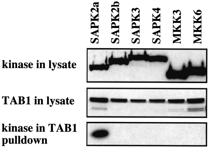
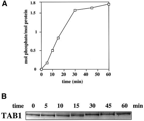
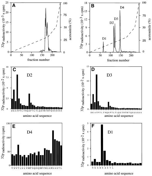
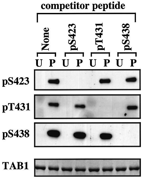
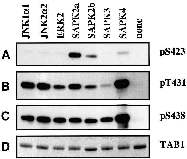
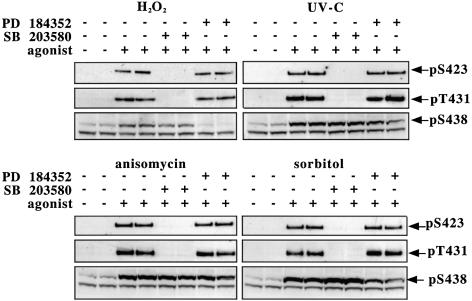
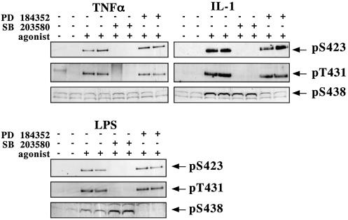
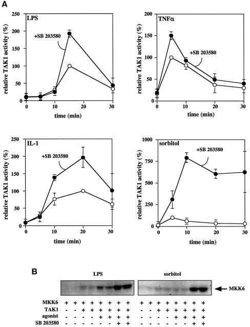
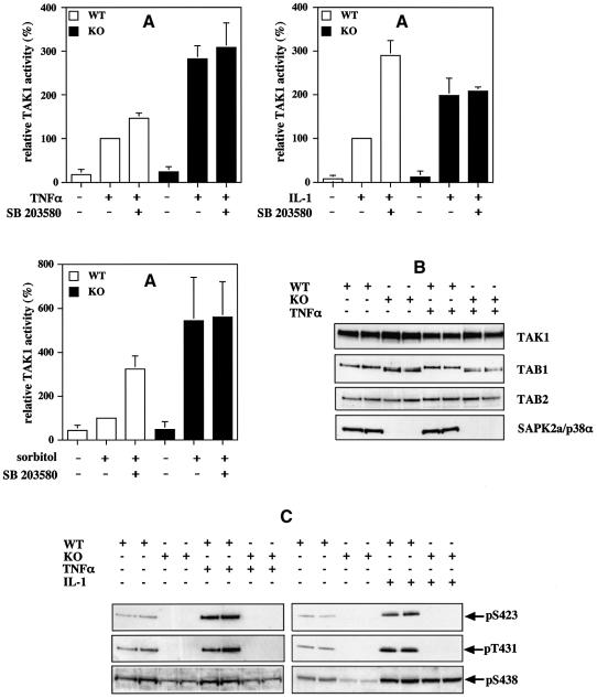
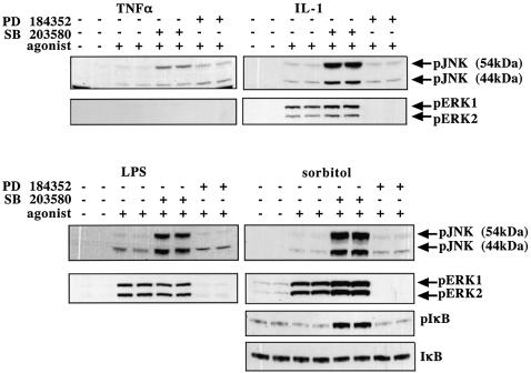
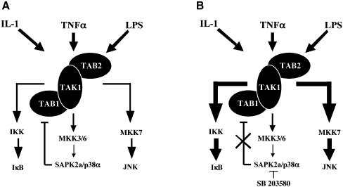
TAB1, a subunit of the kinase TAK1, was phosphorylated by SAPK2a/p38alpha at Ser423, Thr431 and Ser438 in vitro. TAB1 became phosphorylated at all three sites when cells were exposed to cellular stresses, or stimulated with tumour necrosis factor-alpha (TNF-alpha), interleukin-1 (IL-1) or lipopolysaccharide (LPS). The phosphorylation of Ser423 and Thr431 was prevented if cells were pre-incubated with SB 203580, while the phosphorylation of Ser438 was partially inhibited by PD 184352. Ser423 is the first residue phosphorylated by SAPK2a/p38alpha that is not followed by proline. The activation of TAK1 was enhanced by SB 203580 in LPS-stimulated macrophages, and by proinflammatory cytokines or osmotic shock in epithelial KB cells or embryonic fibroblasts. The activation of TAK1 by TNF-alpha, IL-1 or osmotic shock was also enhanced in embryonic fibroblasts from SAPK2a/p38alpha-deficient mice, while incubation of these cells with SB 203580 had no effect. Our results suggest that TAB1 participates in a SAPK2a/p38alpha-mediated feedback control of TAK1, which not only limits the activation of SAPK2a/p38alpha but synchronizes its activity with other signalling pathways that lie downstream of TAK1 (JNK and IKK).


Figures

None
Fig. 1. TAB1 interacts specifically with SAPK2a/p38α. HEK 293 cells were transfected with plasmids coding for GST–TAB1 and SAPK2a/p38α, SAPK2b/p38β2, SAPK3/p38γ, SAPK4/p38δ, MKK3 or MKK6 fused to an HA epitope tag. After 36 h, the cells were lysed and the cell lysate (20 µg of protein) denatured in SDS, subjected to SDS–PAGE, transferred onto nitrocellulose membrane and the membrane probed with an antibody specific for the HA epitope to detect the expression of each protein kinase or with an anti-GST antibody to detect the level of expression of GST–TAB1. The GST–TAB1 in the cell lysates was then affinity purified on glutathione–Sepharose and proteins binding to this support were subjected to SDS–PAGE and immunoblotting with the anti-HA antibody to investigate the binding of each protein kinase to TAB1.
None
Fig. 2. TAB1 is phosphorylated stoichiometrically by SAPK2a/p38α. Bacterially expressed His-TAB1 was phosphorylated at 30°C with 2 U/ml GST–SAPK2a/p38α and Mg [γ-32P]ATP. At the times indicated, an aliquot of the reaction was denatured in SDS and subjected to SDS–PAGE. The gel was stained with Coomassie blue and destained (B). The band corresponding to His-TAB1 was excised and analysed by Cerenkov counting to quantitate the incorporation of phosphate into TAB1 (A).
None
Fig. 3. Identification of the residues on TAB1 phosphorylated by SAPK2a/p38α. His-TAB1 was phosphorylated with active GST–SAPK2a/p38α and subjected to SDS–PAGE. The band corresponding to 32P-labelled TAB1 was visualized by staining with Coomassie blue, excised and digested with trypsin. (A) The tryptic phosphopeptides were separated by HPLC on a Vydac C18 column equilibrated in 0.1% (v/v) trifluoroacetic acid. The column was developed with an acetonitrile gradient in 0.1% (v/v) trifluoroacetic acid (broken line). Radioactivity is indicated by the full line. (B) All the major 32P-labelled peptides from (A) were pooled, further digested with the protease Asp-N and rechromatographed on the Vydac C18 column as in (A). Peptides D2, D3, D4 and D1 were then subjected to solid phase sequencing in (C), (D), (E) and (F), respectively, to determine the sites of phosphorylation. The methodology has been described previously (Stokoe et al., 1992).
None
Fig. 4. Development of phospho-specific antibodies recognizing each of the three phosphorylation sites on TAB1. Bacterially expressed His-TAB1 (0.5 µg) was phosphorylated with SAPK2a/p38α (P) or left unphosphorylated (U). The proteins were then separated by SDS–PAGE, transferred to nitrocellulose and probed with each of the three phospho-specific antibodies in the absence (none) or presence of 10 µg/ml of one of the three phosphopeptide immunogens (termed competitor peptide). Each antibody also contained 10 µg/ml of the unphosphorylated form of the relevant peptide immunogen to neutralize any antibodies present that recognize the unphosphorylated species. The bottom panel shows the amount of His-TAB1 in each gel lane stained with Coomassie Blue.
None
Fig. 5. Phosphorylation of TAB1 in vitro by different MAP kinase family members. Bacterially expressed His-TAB1 (0.5 µg) was phosphorylated with the protein kinases indicated, subjected to SDS-–PAGE and the proteins either transferred onto a nitrocellulose membrane (AC) or stained with Coomassie blue (D). They were then immunoblotted with antibodies recognizing phosphorylated Ser423, Thr431 or Ser438 as in Figure 6.
None
Fig. 6. TAB1 is phosphorylated on Ser423, Thr431 and Ser438 in response to stressors. KB cells were pre-treated for 1 h with or without 10 µM SB 203580 or 2 µM PD184352 then stimulated with H2O2 (2 mM for 15 min), UV-C (30 min), anisomycin (10 µg/ml for 30 min) or sorbitol (0.5 M for 30 min) and lysed. For anti-phospho-Ser423 (pS423) and anti-phospho-Thr431 (pT431) blots, 2 µg of an antibody that immunoprecipitates TAB1 was incubated for 1 h at 4°C with 250 µg of protein lysate. The immunocomplex was collected and washed as described in Materials and methods. The immunoprecipitated proteins were then denatured in SDS, separated by SDS–PAGE, transferred to a nitrocellulose membrane and blotted with phospho-specific antibodies that recognize TAB1 phosphorylated at Ser423 or Thr431. The antibody that recognizes TAB1 phosphorylated at Ser438 (pS438) was more sensitive and immunoblotting could be carried out using the cell lysate without immunoprecipitation. A protein migrating slightly faster than TAB1 was also recognized non-specifically by the anti-pSer438 antibody.
None
Fig. 7. TAB1 is phosphorylated on Ser423, Thr431 and Ser438 in LPS-stimulated RAW macrophages and TNF-α- or IL-1-stimulated KB cells. Cells were pre-treated for 1 h with or without 10 µM SB 203580 or 2 µM PD184352 then stimulated with LPS (100 ng/ml for 15 min), TNF-α (50 ng/ml for 5 min) or IL-1α (20 ng/ml for 20 min), and lysed and analysed as described in the legend to Figure 6.
None
Fig. 8. SB 203580 enhances the activity of TAK1 in LPS-stimulated RAW macrophages and TNF-α-, IL-1- or sorbitol-stimulated KB cells. (A) Cells were pre-treated for 1 h with or without 10 µM SB 203580 then stimulated with LPS (RAW 264.7 macrophages), TNF-α, IL-1 or sorbitol (KB cells). The cells were lysed at the times indicated and the TAK1 complexed to TAB1 was immunoprecipitated from 150 µg of protein lysate using 2 µg of a TAB1-specific antibody bound to protein G–Sepharose. The immune complex was then assayed for TAK1 activity as described in Materials and methods. The results are expressed as the mean ± SEM for three separate experiments with all assays performed in duplicate. Results are shown as a percentage of the activity of TAK1 measured at the time at which activation was maximal in the absence of SB 203580. (B) Same as (A), except that the TAB1 immunoprecipitates were assayed for TAK1 activity by the phosphorylation of MKK6 as described in Materials and methods. The figure shows the incorporation of 32P into the MKK6 substrate as revealed by autoradiography.
None
Fig. 9. The TNF-α-induced activation of TAK1 is enhanced in embryonic mouse fibroblasts deficient in SAPK2a/p38α. Immortalized embryonic fibroblasts from wild-type (WT, open bars) and SAPK2a/p38α knockout (KO, black bars) mice were treated for 1 h with or without 10 µM SB 203580, then stimulated for 10 min with 10 ng/ml of mouse TNF-α, for 20 min with 20 ng/ml of mouse IL-1 or for 30 min with 0.5 M sorbitol, and the cells lysed. (A) The complexes containing TAK1 were immunoprecipitated from the cell lysates (0.15 mg of protein) using a TAB1-specific antibody, and the activity of TAK1 in the cell lysates determined (see Materials and methods). The results are expressed as the mean ± SEM for three separate experiments with all assays performed in duplicate. Results are shown as a percentage of the activity of TAK1 in stimulated wild-type cells measured without pre-incubation with SB 203580. (B) The TAK1 complexes immunoprecipitated from the lysates of cells stimulated with TNF-α were prepared as in (A), then subjected to SDS–PAGE followed by immunoblotting with antibodies that recognize TAK1, TAB1 and TAB2. Cell lysate (20 µg of protein) was also subjected to SDS–PAGE followed by immunoblotting for SAPK2a/p38α (C). TAK1 complexes immunoprecipitated from the lysates of cells stimulated with TNF-α or IL-1 were prepared as in (A), and then analysed as in (B), except that the gels were immunoblotted with antibodies that recognize TAB1 phosphorylated at Ser423, Thr431 or Ser438.
None
Fig. 10. SB 203580 enhances the activation of JNK isoforms and phosphorylation of IκB. KB cells were stimulated with 50 ng/ml TNF-α (5 min), 20 ng/ml IL-1 (20 min) or 0.5 M sorbitol (30 min), and RAW264 macrophages were stimulated with 100 ng/ml LPS (15 min). The cells were incubated for 1 h without (–) or with (+) 2 µM PD 184352 or 10 µM SB 203580 prior to stimulation with each agonist. The cells were lysed and lysate protein (30 µg) was subjected to SDS–PAGE and immunoblotted with phospho-specific antibodies that recognize the active forms of JNK isoforms, the active forms of ERK1 and ERK2, IκB phosphorylated at Ser32, and an antibody that recognizes all forms of IκB.
None
Fig. 11. Schematic representation of the feedback control of TAK1 activity by SAPK2a/p38α and its implication for the regulation of JNK and NF-κB. (A) SAPK2a/p38α downregulates TAK1, probably via the phosphorylation of TAB1. (B) The inhibition of SAPK2a/p38α by SB 203580 (or other inhibitors of this protein kinase) abolishes this feedback control of TAK1, causing upregulation of the JNK and IKK pathways (illustrated by the thicker arrows).

References

    1. Adams R.H. et al. (2000) Essential role of p38α MAP kinase in placental but not embryonic cardiovascular development. Mol. Cell, 6, 109–116. - PubMed
    1. Alessi D.R., Smythe,C. and Keyse,S.M. (1993) The human CL100 gene encodes a Tyr/Thr-protein phosphatase which potently and specifically inactivates MAP kinase and suppresses its activation by oncogenic ras in Xenopus oocyte extracts. Oncogene, 8, 2015–2020, - PubMed
    1. Alessi D.R., Cohen,P., Ashworth,A., Cowley,S., Leevers,S.J. and Marshall,C.J. (1995) Assay and expression of mitogen-activated protein kinase, MAP kinase kinase and Raf. Methods Enzymol., 255, 279–290. - PubMed
    1. Alonso G., Ambrosino,C., Jones,M. and Nebreda,A.R. (2000) Differential activation of p38 mitogen-activated protein kinase isoforms depending on signal strength. J. Biol. Chem., 275, 40641–40648. - PubMed
    1. Ambrosino C., Mace,G., Galban,S., Fritsch,C., Vintersten,K., Black,E., Gorospe,M. and Nebreda,A.R. (2002) Negative feedback regulation of MKK6 mRNA stability by p38α mitogen-activated protein kinase. Mol. Cell. Biol., 23, 370–381. - PMC - PubMed

Publication types

MeSH terms

Substances