. 2003 Nov 3;22(21):5851-62.
doi: 10.1093/emboj/cdg553.

A chromosomal SIR2 homologue with both histone NAD-dependent ADP-ribosyltransferase and deacetylase activities is involved in DNA repair in Trypanosoma brucei

Affiliations

A chromosomal SIR2 homologue with both histone NAD-dependent ADP-ribosyltransferase and deacetylase activities is involved in DNA repair in Trypanosoma brucei

José A García-Salcedo et al. EMBO J. .

Abstract

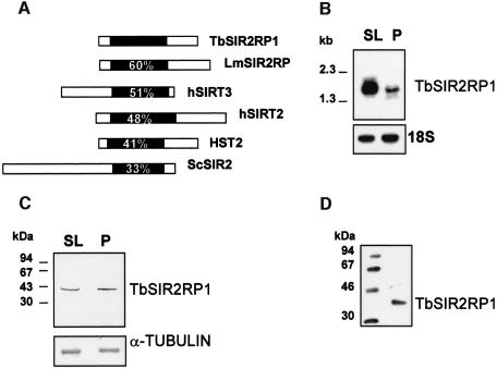
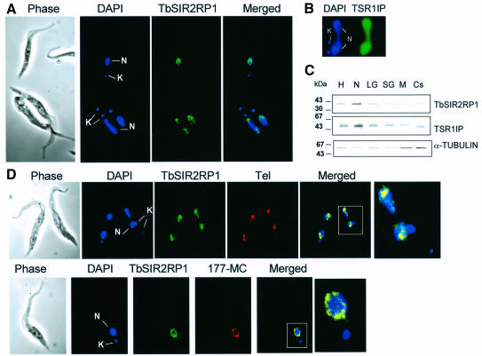
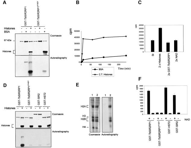
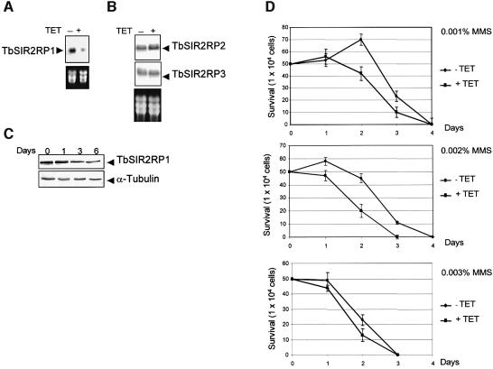
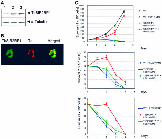
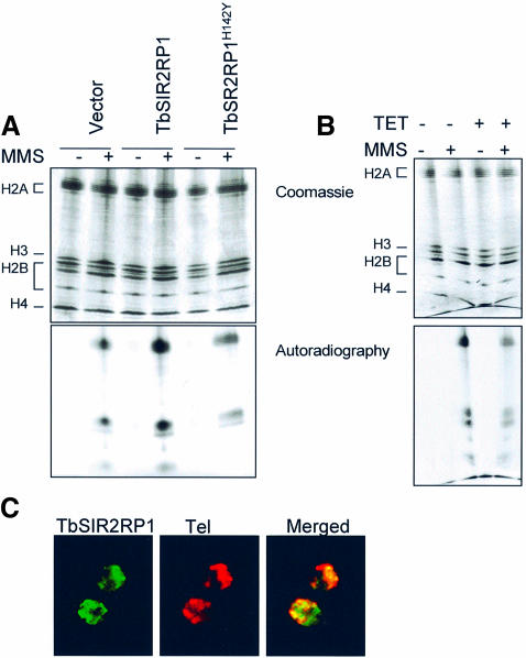
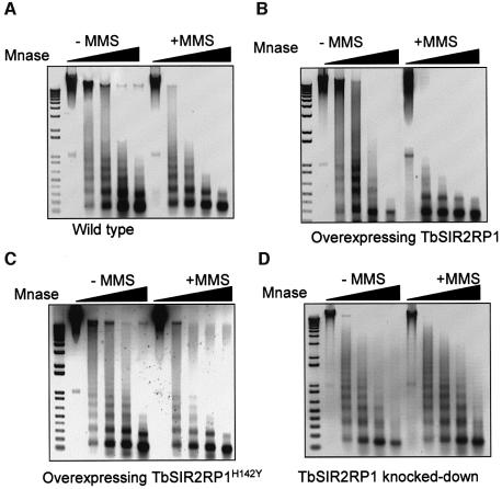
SIR2-like proteins have been implicated in a wide range of cellular events including chromosome silencing, chromosome segregation, DNA recombination and the determination of life span. We report here the molecular and functional characterization of a SIR2-related protein from the protozoan parasite Trypanosoma brucei, which we termed TbSIR2RP1. This protein is a chromosome-associated NAD-dependent enzyme which, in contrast to other known proteins of this family, catalyses both ADP-ribosylation and deacetylation of histones, particulary H2A and H2B. Under- or overexpression of TbSIR2RP1 decreased or increased, respectively, cellular resistance to DNA damage. Treatment of trypanosomal nuclei with a DNA alkylating agent resulted in a significant increase in the level of histone ADP-ribosylation and a concomitant increase in chromatin sensitivity to micrococcal nuclease. Both of these responses correlated with the level of TbSIR2RP1 expression. We propose that histone modification by TbSIR2RP1 is involved in DNA repair.


Figures

None
Fig. 1. Characterization of TbSIR2RP1. (A) Sequence comparison between TbSIR2RP1 and homologous proteins. The position of the conserved core domain is shown in black, and numbers represent the percentage amino acid identity with this region of TbSIR2RP1. The SIR2-like proteins compared are: LmSIR2RP from L.major (Yahiaoui et al., 1996), hSIRT2 and hSIRT3 from man (Frye, 1999) and HST2 and SIR2 from S.cerevisiae (Brachmann et al., 1995). (B) Northern blot analysis of TbSIR2RP1 mRNA. A 10 µg aliquot of poly(A)+ RNA from T.brucei AnTat 1.1 bloodstream long slender (SL) and procyclic forms (P) was separated on a formaldehyde agarose gel, transferred to nitrocellulose and probed with 32P-labelled TbSIR2RP1 cDNA. The filter was subsequently rehybridized with a ribosomal 18S probe, as control for RNA loading. (C) Immunoblot analysis of TbSIR2RP1. Total bloodstream slender (SL) and procyclic (P) cell lysates in SDS–PAGE sample buffer were examined by western blot analysis with affinity- purified anti-TbSIR2RP1. A control analysis was performed on a similar filter with an anti-α-tubulin monoclonal antibody. (D) Immunoprecipitation of in vitro translated TbSIR2RP1 with affinity-purified anti-TbSIR2RP1 antibodies.
None
Fig. 2. Cellular localization of TbSIR2RP1. (A) Procyclic forms were stained by immunofluorescence with anti-TbSIR2RP1 antibodies and counterstained with DAPI. The DAPI staining reveals the location of the nucleus (N) and kinetoplast (mitochondrial DNA, K). (B) Immunolocalization of TSR1IP nuclear protein. (C) Distribution of TbSIR2RP1 in subcellular fractions from procyclic forms of T.brucei. Subcellular fractions were separated by SDS–PAGE, transferred to nitrocellulose and probed with anti-TbSIR2RP1, anti-α-tubulin and anti-TSR1IP antibodies. Subcellular fraction abbreviations are as follows: H, homogenate; N, nuclear; LG, large granular (mitochondrial); SG, small granular (glycosomal); M, endosomal; Cs, cytoplasmic (soluble). (D) TbSIR2RP1 co-localizes with telomeres and minichromosomes. Procyclic forms were fixed and subjected to IF and FISH using anti-TbSIR2RP1 antibodies and probes for telomeric repeats (Tel) and minichromosome (177-MC). Overlap of the two signals is yellow/orange.
None
Fig. 3. NAD-dependent ribosylation and deacetylation of proteins by TbSIR2RP1. (AIn vitro ribosylation reactions were performed with 1 µg of GST–TbSIR2RP1 or GST–TbSIR2RP1H142Y, plus 5 µg of BSA and 5 µg of histones using [32P]NAD as donor. The top panel illustrates a Coomassie-stained gel of reaction products resolved by 10% SDS–PAGE, whereas the bottom panel shows the autoradiograph of the gel. (B) Time curves of histone and BSA ribosylation by GST–TbSIR2RP1 at 37°C. Each point represents the ribosylation products of 5 µg of substrate by 0.5 µg of GST–TbSIR2RP1 using 5 µCi of [32P]NAD at the indicated time. Reaction products were precipitated with 20% TCA (w/v), collected and washed on a GF/C glass fibre filter (Whatman), and then counted after addition of liquid scintillation fluid. (C) Ribosylation reactions were performed with 5 µg of histones, 0.5 µg of GST–TbSIR2RP1 and 5 µCi of [32P]NAD (standard reaction, column St) or a double concentration of histone (10 µg, column 2× histones), GST–TbSIR2RP1 (1 µg, column 2× GST–TbSIR2RP1) and [32P]NAD (10 µCi, column 2× NAD). (D) Comparison of TbSIR2RP1 ribosylation activity with that of other members of the SIR2-like family. Ribosylation reactions with 1 µg of GST–TbSIR2RP1, GST–TbSIR2RP1H142Y, GST–SIR2 or GST–HST2 were performed with and without 5 µg of histones using [32P]NAD as donor. The top panel illustrates a Coomassie-stained gel of the reaction products, and the bottom panel shows the autoradiograph of the gel. (E) Triton–acid–urea (TAU) gel of T.brucei histones treated with GST–TbSIR2RP1 or GST–TbHST1H142Y and [32P]NAD, respectively stained with Coomassie blue and autoradiographed. The histone types are indicated on the left. (F) Analysis of the NAD-dependent histone deacetylase activity. Histones were acetylated with [3H]acetyl-CoA by HAT1 and then treated with GST–TbSIR2RP1, GST– TbSIR2RP1H142Y, GST–HST2 and GST–SIR2. The amount of acetate released was measured in the presence or absence of NAD.
None
Fig. 4. Effect of TbSIR2RP1 RNAi on cell sensitivity to DNA damage. (A) Northern blot analysis of mRNA levels from cells incubated for 24 h without (–) or with (+) tetracycline. rRNAs stained with ethidium bromide are shown as loading controls. (B) A similar northern blot was hybridized with probes corresponding to the entire ORF of two other trypanosomal homologues of SIR2 (TbSIR2RP2, accession No. AC119406; TbSIR2RP3, accession No. AF102869). (C) Western blot analysis of whole-cell lysate (1 × 106 cell equivalents/lane) from uninduced (day 0) and tetracycline- induced cells (days 1–6) using anti-TbSIR2RP1 and anti-α-tubulin antibodies. (D) Cell survival curves are shown for uninduced (TET–) and tetracycline-induced cells (TET+) treated with different concentrations of MMS.
None
Fig. 5. Effect of ectopic expression of TbSIR2RP1 and TbSIR2RP1H142Y on cell sensitivity to MMS. (A) Western blot analysis of control cells (lane 1) or cells overexpressing TbSIR2RP1 (lane 2) and TbSIR2RP1H142Y (lane 3) using anti-TbSIR2RP1 and anti-α-tubulin antibodies. (B) Subcellular localization of TbSIR2RP1 in cells overexpressing the protein. Procyclic forms overexpressing TbSIR2RP1 were subjected to IF and FISH using anti-TbSIR2RP1 antibodies and probes for telomeric repeats (Tel). Overlap of the two signals is yellow/orange. (C) Cell survival in the presence of different concentrations of MMS, comparing wild-type and cells overexpressing TbSIR2RP1 and TbSIR2RP1H142Y.
None
Fig. 6. Extent of histone ribosylation in response to DNA damage correlates with the cellular concentration of TbSIR2RT1. The results of TAU gel analysis of histone ribosylation in nuclei from trypanosomes overexpressing TbSIR2RP1 and TbSIR2RP1H142Y (A) and from TbSIR2RP1 knocked-down cells (B). Histones were purified from nuclei treated for 1 h with MMS and [32P]NAD in the presence of 5 mM 3-aminobenzamide. The bottom panel shows the autoradiograph of the gel. (C) The localization of TbSIR2RP1 in cells with damaged DNA. Procyclic forms incubated in 0.001% MMS for 12 h were subjected to IF and FISH with anti-TbSIR2RP1 antibodies and telomeric probes, respectively.
None
Fig. 7. Alkylated chromatin is more sensitive to MNase. Untreated and MMS-treated chromatin from wild-type procyclic cells (A) and from cell overexpressing TbSIR2RP1 (B) or TbSIR2RP1H142Y (C), or from TbSIR2RP1 knocked-down cells (D) were incubated with MNase. All digestion reactions contained 4 µg of DNA. Chromatin was digested with 0.00025, 0.0005, 0.0015 and 0.005 U of MNase for 10 min at 30°C. A negative print of the ethidium-stained DNA is shown. A 1 kb PLUS DNA ladder (Invitrogen) is on the left of each panel.

References

    1. Adamietz P. and Rudolph,A. (1984) ADP-ribosylation of nuclear proteins in vivo. Identification of histone H2B as a major acceptor for mono- and poly(ADP-ribose) in dimethyl sulfate-treated hepatoma AH 7974 cells. J. Biol. Chem., 259, 6841–6846. - PubMed
    1. Afshar G. and Murnane,J.P. (1999) Characterization of a human gene with sequence homology to Saccharomyces cerevisiae SIR2. Gene, 234, 161–168. - PubMed
    1. Bell S.D., Botting,C.H., Wardleworth,B.N., Jackson,S.P. and White,M.F. (2002) The interaction of Alba, a conserved archaeal chromatin protein, with Sir2 and its regulation by acetylation. Science, 296, 148–151. - PubMed
    1. Bohm L., Schneeweiss,F.A., Sharan,R.N. and Feinendegen,L.E. (1997) Influence of histone acetylation on the modification of cytoplasmic and nuclear proteins by ADP-ribosylation in response to free radicals. Biochim. Biophys Acta, 1334, 149–154. - PubMed
    1. Borra M.T., O’Neill,F.J., Jackson,M.D., Marshall,B., Verdin,E., Foltz,K.R. and Denu,J.M. (2002) Conserved enzymatic production and biological effect of O-acetyl-ADP-ribose by silent information regulator 2-like NAD+-dependent deacetylases. J. Biol. Chem., 277, 12632–12641. - PubMed

Publication types