. 2003 Nov 25;100(24):14127-32.
doi: 10.1073/pnas.2233012100. Epub 2003 Oct 30.

Exploring the functional complexity of cellular proteins by protein knockout

Affiliations

Exploring the functional complexity of cellular proteins by protein knockout

Jianxuan Zhang et al. Proc Natl Acad Sci U S A. .

Abstract

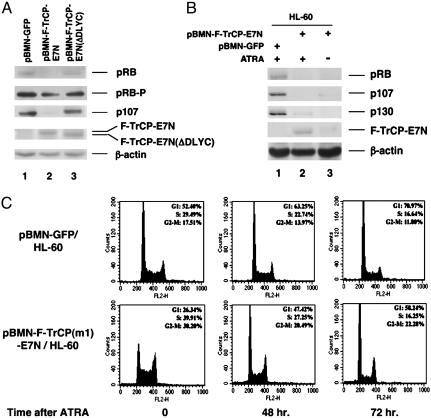
Comprehensive dissection of protein functions entails more complicated manipulations than simply eliminating the protein of interest. Established knockdown technologies, such as RNA interference, antisense oligodeoxynucleotides, or ribozymes, are limited for specific applications such as modulating protein levels or specific targeting of a posttranslationally modified subpopulation. Here we show that the engineered Skp1, Cullin 1, and F-box-containing betaTrCP substrate receptor ubiquitin-proteolytic system, designated protein knockout, could achieve not only total elimination but also rapid and systematic reduction of a given cellular protein. Stable expression of a single engineered betaTrCP demonstrated simultaneous and sustained degradation of the entire retinoblastoma family proteins. Furthermore, the engineered betaTrCP was capable of selecting hypo- but not hyperphosphorylated forms of retinoblastoma for degradation. The engineered betaTrCP has been extensively modified to increase its specificity in substrate selection. This optimized protein-knockout system offers a powerful and versatile proteomic tool to dissect diverse functional properties of cellular proteins in somatic cells.


Figures

Fig. 1.
Fig. 1.
Systematic reduction of the endogenous p107 by F-TrCP-E7N. C33A cells were infected with increasing doses of recombinant Ad-F-TrCP-E7N adenovirus for 48 h. The levels of endogenous p107, cyclin A, Cdk2, and β-actin (loading control) were detected by immunoblotting with the respective antibodies, and in comparison with the dose-dependent increase in the expression of F-TrCP-E7N. The percentages of p107 remaining in each adenovirus-infected cell were quantitated by densitometry scanning and are indicated.
Fig. 2.
Fig. 2.
Selective degradation of hypophosphorylated form of RB in U2OS cells. (A) U2OS cells contain both hypophosphorylated and hyperphosphorylated forms of RB. Immunoblotting was carried out to detect RB in U2OS cell extracts by using antibodies that recognize both RB forms (lane 1) (IF8, Santa Cruz Biotechnology), or specific for hypo- (lane 2) (G99–549, BD Biosciences) or hyperphosphorylated RB (lane 3) [(phospho-RB (Ser 807/811) polyclonal antibody, Cell Signaling]. (B) U2OS cells were infected with increasing doses (in μl) of Ad1-F-TrCP-E7N or control pAd1 adenoviruses for 48 h. Cell extracts were prepared and immunoblotted with antibodies against RB, p107, FLAG (M2), p21waf1, p27kip1, and β-actin.
Fig. 3.
Fig. 3.
Structure-based mutagenesis of the substrate-binding residues on the engineeredβTrCP-E7N ubiquitin–protein ligase. (A) Schematic diagrams of the predicted SCFβTrCP-BP/IκB/target and SCFβTrCP(m1)-BP/target complexes. BP, binding peptide; T, target. (B) Three-dimensional model of the WD40 repeats of βTrCP. The five positively charged residues are cyan. (C) The modified F-TrCP(m1)-E7N ubiquitin–protein ligase cannot bind to IκB, but retains its interaction with p107. HeLa cells were transiently transfected with IκB, together with pcDNA3 (lane 1), F-TrCP (lane 2), F-TrCP-E7N (lane 3), and F-TrCP(m1)-E7N (lane 4), and treated with MG132 for2hand then with TNF-α for 15 min. Cell extracts were prepared for immunoprecipitation with the anti-FLAG (M2) antibody, and probed with either phosphorylated IκB antibody or anti-p107 polyclonal antibody. F-TrCP(m1)-E7N migrates as a doublet on SDS gels for reasons yet to be determined (lane 4). (D)Efficient degradation of p107 by F-TrCP(m1)-E7N in C33A cells. C33A cells transfected with the indicated plasmids and 1 μg of pCMV-CD19 were enriched by immunomagnetic selection, and levels of endogenous p107 were examined by immunoblotting. (E) Indirect immunofluorescence assay was carried out to detect endogenous p107 (Center) in response to transient transfection and expression of F-TrCP, F-TrCP-E7N, or F-TrCP(m1)-E7N (Left) in individual C33A cells (marked by arrows). Images of the same field of cells counterstained with 4′,6-diamidino-2-phenylindole (DAPI) were shown to identify the nuclei of both transfected (marked by arrows) and untransfected cells.
Fig. 4.
Fig. 4.
The minimal p107-binding domain of E7 is sufficient to recruit p107 for targeted destruction. (A) The engineered F-TrCP-10GS-E7m ubiquitin–protein ligase efficiently binds to p107. C33A cells transiently transfected with the indicated plasmids were treated with the proteasome inhibitor MG132 for 2 h. Cell extracts were prepared for immunoprecipitation with the anti-FLAG antibody, and were probed with antibodies against FLAG or p107. (B) Degradation of the endogenous p107 by the engineered F-TrCP-E7m. C33A cells were transiently transfected with the indicated plasmids (in μg) and 1 μg of pCMV-CD19. Transfected cells were enriched by immunomagnetic bead selection, and the steady-state levels of p107, F-TrCP fusions, and β-actin (loading control) were determined by immunoblotting using antibodies against p107, FLAG, and β-actin. The experiment was repeated four times, and the amount of p107 remaining was measured by PhosphorImager analysis. Endogenous β-actin levels were also measured as an internal loading control.
Fig. 5.
Fig. 5.
Retroviral-mediated delivery of the engineered F-TrCP-E7N ubiquitin–protein ligase for targeted degradation of RB family proteins in the HL-60 cells. (A) HL-60 cells infected with the indicated recombinant retroviruses were isolated by FACS, and the steady state levels of RB, p107, F-TrCP-E7N, and F-TrCP-E7N(ΔDLYC) were determined by immunoblotting using the respective antibodies. β-Actin levels were also determined as a specificity and internal loading control. RB and RB-P denote hypo- and hyperphosphorylated RB. (B) Infected and FACS-sorted HL-60 cells were cultured for 3 months, and either untreated (lane 3) or treated with 1 μM of ATRA for 72 h (lane 2). Endogenous RB, p107, and p130 were determined in response to the expression of F-TrCP-E7N by immunoblotting. (C) FACS analysis to determine the DNA contents of the live HL-60 cells infected by control or pBMN-F-TrCP(m1)-E7N retroviruses under normal growth conditions and in response to ATRA-induced growth arrest.

References

    1. Zhou, P., Bogacki, R., McReynolds, L. & Howley, P. M. (2000) Mol. Cell 6, 751–756. - PubMed
    1. Hershko, A. & Ciechanover, A. (1998) Annu. Rev. Biochem. 67, 425–479. - PubMed
    1. Yaron, A., Hatzubai, A., Davis, M., Lavon, I., Amit, S., Manning, A. M., Andersen, J. S., Mann, M., Mercurio, F. & Ben-Neriah, Y. (1998) Nature 396, 590–594. - PubMed
    1. Winston, J. T., Strack, P., Beer-Romero, P., Chu, C. Y., Elledge, S. J. & Harper, J. W. (1999) Genes. Dev. 13, 270–283. - PMC - PubMed
    1. Tan, P., Fuchs, S. Y., Chen, A., Wu, K., Gomez, C., Ronai, Z. & Pan, Z. Q. (1999) Mol. Cell 3, 527–533. - PubMed

Publication types

MeSH terms

LinkOut - more resources