. 2003 Nov 11;100(23):13495-500.
doi: 10.1073/pnas.2334503100. Epub 2003 Oct 30.

Generation of hair cells by stepwise differentiation of embryonic stem cells

Affiliations

Generation of hair cells by stepwise differentiation of embryonic stem cells

Huawei Li et al. Proc Natl Acad Sci U S A. .

Abstract

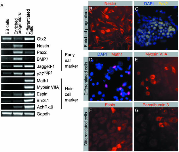
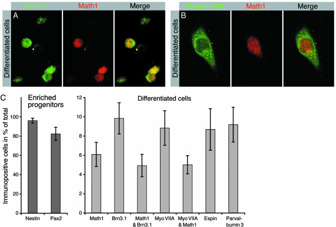
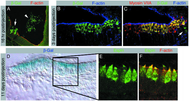
The increase in life expectancy is accompanied by the growing burden of chronic diseases. Hearing loss is perhaps the most prevalent of all chronic diseases. In addition to age-related hearing loss, a substantial number of cases of audiological impairment are either congenital in nature or acquired during childhood. The permanence of hearing loss is mainly due to the inability of the cochlear sensory epithelium to replace lost mechanoreceptor cells, or hair cells. Generation of hair cells from a renewable source of progenitors that can be transplanted into damaged inner ears is a principal requirement for potential cell replacement therapy in this organ. Here, we present an experimental protocol that enables us to routinely create inner ear progenitors from murine embryonic stem cells in vitro. These progenitors express a comprehensive set of marker genes that define the developing inner ear, in particular the organ's developing sensory patches. We further demonstrate that cells that express markers characteristic of hair cells differentiate from embryonic stem cell-derived progenitors. Finally, we show that these progenitors integrate into the developing inner ear at sites of epithelial injury and that integrated cells start expressing hair cell markers and display hair bundles when situated in cochlear or vestibular sensory epithelia in vivo.


Figures

Fig. 1.
Fig. 1.
Analysis of expression of markers by ES cells, in vitro selectively enriched progenitor cells, and cells after in vitro differentiation. (A) RT-PCR-based analysis of expression of marker transcripts. Enriched progenitor cells were analyzed after expansion with bFGF. Differentiated cells were analyzed after 14 days of in vitro differentiation. Expression analysis of the ubiquitously expressed GAPDH is shown for reference. (B) Virtually all selected progenitors express nestin. (C) The majority of cells display Pax2 immunoreactivity. Nuclei are visualized with blue 4,6-diamidino-2-phenylindole staining. (D) After 14 days of in vitro differentiation, nuclear immunoreactivity for the hair cell marker Math1 was detectable. (E) A subset of differentiated cells express the hair cell marker myosin VIIA. (F) The hair-bundle marker espin is detectable in differentiated cells. (G) The hair cell marker parvalbumin 3 is detectable in differentiated cells.
Fig. 2.
Fig. 2.
Coexpression of hair cell markers by differentiated cells. (A) Expression of the transcription factors Brn3.1 and Math1 in nuclei of differentiated cells. The majority of Math1-expressing cells coexpress Brn3.1. (B) A large fraction of Math1-expressing cells coexpress the hair cell marker protein myosin VIIA. (C) Quantification of the number of cells expressing otic vesicle and hair cell markers at the progenitor cell stage (dark gray bars) and after in vitro differentiation (light gray bars). The individual bars visualize the fraction of immunopositive cells of the total number of cells. Shown are mean values and standard deviations determined in three to seven independent experiments for each data set.
Fig. 3.
Fig. 3.
Grafted inner ear progenitor cells integrate into the developing inner ear and differentiate into hair cells. (A) Cross section of the developing chicken inner ear at embryonic day 3.5, 16 h postinjection of ROSA26-derived progenitor cells. β-gal-expressing murine cells invade the epithelium that surrounds the lumen of the otic vesicle (arrow). The arrowhead labels β-gal-positive murine cells that remain in the lumen of the otic vesicle. F-actin labeling reveals disruptions of the epithelium, caused by injury during the grafting procedure. (B-C) Longitudinal section of a developing cochlear sensory epithelium at embryonic day 6, 3 days postinjection, immunolabeled with antibodies for β-gal (B) and myosin VIIA (C). F-actin labeling is shown for orientation. (B) β-gal-positive cells occur in the developing sensory patch. (C) Myosin VIIA-positive early hair cells can be detected in the cochlea at embryonic day 6. Merging the confocal images reveals a patch of myosin VIIA-positive hair cells that express β-gal. β-gal-positive cells observed in the developing supporting cell layer, located below the apical cell layer of early hair cells, do not express myosin VIIA (arrow). (D) At day 14 of embryonic development, 11 days postinjection, we found patches of β-gal-positive cells in inner ear sections. The patch of β-gal-positive cells in a utricular section, here revealed with X-gal histochemistry, is surrounded by cells that did not display blue X-gal staining, which served as negative control for the staining method. (E) Enlargement of an area with β-gal-positive cells exposes espin-positive cells with cylindrical hair cell morphology that display espin-positive hair bundles. (F) Espin-positive hair bundles are rich in F-actin, here apparent as yellow staining in the merged confocal images.

References

    1. Lee, S. H., Lumelsky, N., Studer, L., Auerbach, J. M. & McKay, R. D. (2000) Nat. Biotechnol. 18, 675-679. - PubMed
    1. Lumelsky, N., Blondel, O., Laeng, P., Velasco, I., Ravin, R. & McKay, R. (2001) Science 292, 1389-1394. - PubMed
    1. Doetschman, T. C., Eistetter, H., Katz, M., Schmidt, W. & Kemler, R. (1985) J. Embryol. Exp. Morphol. 87, 27-45. - PubMed
    1. Wichterle, H., Lieberam, I., Porter, J. A. & Jessell, T. M. (2002) Cell 110, 385-397. - PubMed
    1. Pickles, J. O. & van Heumen, W. R. (1997) Dev. Neurosci. 19, 476-487. - PubMed

Publication types

Substances

LinkOut - more resources