. 2003 Nov 11;100(23):13298-302.
doi: 10.1073/pnas.2336149100. Epub 2003 Oct 30.

Src kinase activation by direct interaction with the integrin beta cytoplasmic domain

Affiliations

Src kinase activation by direct interaction with the integrin beta cytoplasmic domain

Elena G Arias-Salgado et al. Proc Natl Acad Sci U S A. .

Abstract

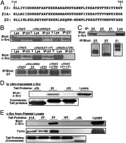
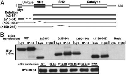
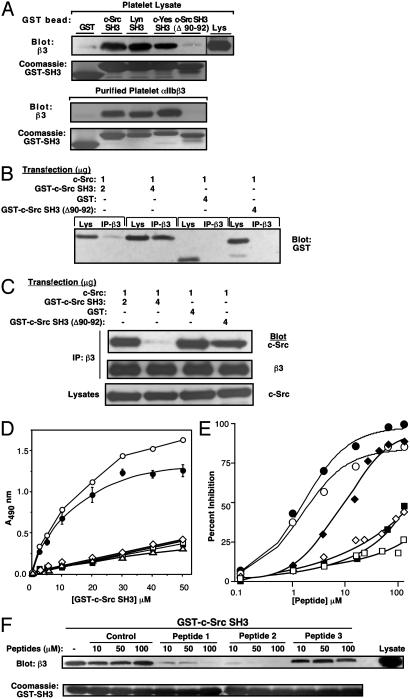
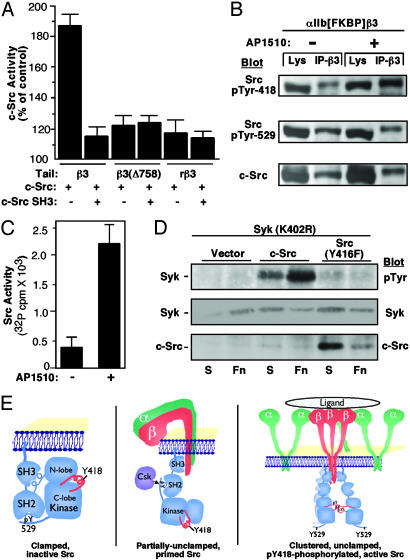
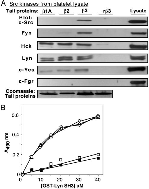
Src tyrosine kinases transmit integrin-dependent signals pivotal for cell movement and proliferation. Here, we establish a mechanism for Src activation by integrins. c-Src is shown to bind constitutively and selectively to beta3 integrins through an interaction involving the c-Src SH3 domain and the carboxyl-terminal region of the beta3 cytoplasmic tail. Clustering of beta3 integrins in vivo activates c-Src and induces phosphorylation of Tyr-418 in the c-Src activation loop, a reaction essential for adhesion-dependent phosphorylation of Syk, a c-Src substrate. Unlike c-Src, Hck, Lyn, and c-Yes bind more generally to beta1A, beta2, and beta3 cytoplasmic tails. These results invoke a model whereby Src is primed for activation by direct interaction with an integrin beta tail, and integrin clustering stabilizes activated Src by inducing intermolecular autophosphorylation. The data provide a paradigm for integrin regulation of Src and a molecular basis for the similar functional defects of osteoclasts or platelets from mice lacking beta3 integrins or c-Src.


Figures

Fig. 1.
Fig. 1.
Interaction of c-Src with the integrin β3 cytoplasmic tail. (A) Integrin β tail sequences. Residue numbers shown are for the full-length β3 subunit. (B) c-Src was transfected into CHO cells expressing the indicated integrins. β3 immunoprecipitates from resuspended cells were probed on Western blots with antibodies to c-Src or β3. Lys, lysate. (C) αIIbβ3 CHO cells were cotransfected with c-Src, αM, and β2. Then β1, β2, and β3 immunoprecipitates were probed as indicated. Immunoprecipitation with preimmune serum yielded no specific bands (data not shown). (D and E) Neutravidin beads coated with integrin tail model proteins were incubated with in vitro-translated c-Src (D) or platelet lysate (E). Bound c-Src or talin was detected by Western blotting. rβ3 is a random β3 tail sequence. Tail protein loading was assessed by Coomassie staining.
Fig. 2.
Fig. 2.
Regions of c-Src necessary for interaction with αIIbβ3. (A) Domain structure of murine c-Src and deletion mutants. (B) αIIbβ3 CHO cells were transfected with c-Src or a deletion mutant, and β3 immunoprecipitates were probed as indicated.
Fig. 3.
Fig. 3.
Interaction of c-Src SH3 with the integrin β3 cytoplasmic tail. (A) Platelet lysate or αIIbβ3 purified from platelets was incubated with glutathione-Sepharose beads coated with the indicated GST-SH3 domains. Bound proteins were eluted and probed with antibody to β3. (B and C) αIIbβ3 CHO cells were cotransfected with c-Src and the indicated GST fusion proteins. β3 immunoprecipitates were probed with antibody to GST (B), c-Src, or β3(C). (D) Direct binding of purified GST-c-Src SH3 to integrin β cytoplasmic tail proteins, assessed by ELISA: β3 (○), rβ3 (▪), β3(Δ758) (□), β1A (⋄), and β2 (▵). Specific binding of c-Src SH3 to β3(•). (E) Inhibition of the β3 tail/c-Src SH3 interaction by peptides. Immobilized β3 tail and soluble GST-c-Src SH3 (10 μM) were incubated with the following peptides: Src SH3-selective (LSSRPLPTLPSP) (•), Src family SH3-selective (KGGRSLRPLPPLPPPG) (○), β3 tail residues 722-741 (⋄), β3 748-762 (♦), αIIb 989-1008 (□), and control (KGELRLRNYYYDVV) (▪). Then, binding of GST-c-Src SH3 was detected by ELISA. (F) Inhibition of the αIIbβ3/c-Src SH3 interaction by peptides. Lysate from αIIbβ3 CHO cells was incubated with GST-c-Src SH3 coupled to beads in the presence of KGELRLRNYYYDVV (control), KGGRSLRPLPPLPPPG (peptide 1), LSSRPLPTLPSP (peptide 2), or APTYPPPLPP (peptide 3). β3 bound to beads was detected by immunoblotting.
Fig. 4.
Fig. 4.
The integrin β3 cytoplasmic tail modulates c-Src activity. (A) Specific binding to the β3 cytoplasmic tail increases the activity of purified c-Src. Activity is expressed relative to a control sample containing c-Src and beads not coated with tail proteins. Data represent means ± SEM of three experiments. (B and C) Oligomerization of cellular β3 integrins activates c-Src. CHO cells expressing αIIb(FKBP)β3 were transfected with c-Src and incubated in suspension with or without AP1510. Then, β3 immunoprecipitates were subjected to immunoblotting with antibodies specific for pTyr-418 and pTyr-529 (B)or in vitro Src kinase assay (C) (means ± SEM of three experiments). In three immunoblotting experiments and after normalization for gel loading, AP1510 increased Tyr-418 phosphorylation by 210 ± 38% and decreased Tyr-529 phosphorylation by 47 ± 2%. (D) c-Src, but not Src(Y416F), can promote adhesion-dependent tyrosine phosphorylation of Syk. c-Src, Src(Y416F), or vector DNA was cotransfected with kinase-inactive Syk(K402R) into CHO cells. Cells were maintained in suspension (S) or plated on fibronectin (Fn) for 30 min, and lysates were subjected to immunoblotting, as indicated. (E) Model for Src activation by clustering of β3 integrins, based on current data and recent models for β3 integrins (32, 34, 35) and Src (25).
Fig. 5.
Fig. 5.
Interaction of Src family kinases with integrin β cytoplasmic tail proteins. (A) Neutravidin beads coated with integrin tail model proteins were incubated with platelet lysate. Bound Src kinases were detected by Western blotting. (B) Direct binding of purified GST-Lyn SH3 to integrin β cytoplasmic tail proteins, assessed by ELISA: β1A (⋄), β2(▵), β3(○), rβ1A (□), and rβ3(▪).

Similar articles

Cited by

References

    1. Hynes, R. (2002) Cell 110, 673-687. - PubMed
    1. Lowell, C. A., Fumagalli, L. & Berton, G. (1996) J. Cell Biol. 133, 895-910. - PMC - PubMed
    1. Klinghoffer, R. A., Sachsenmaier, C., Cooper, J. A. & Soriano, P. (1999) EMBO J. 18, 2459-2471. - PMC - PubMed
    1. Parsons, J. T. (2003) J. Cell Sci. 116, 1409-1416. - PubMed
    1. Hruska, K. A., Rolnick, F., Huskey, M., Alvarez, U. & Cheresh, D. (1995) Endocrinology 136, 2984-2992. - PubMed

Publication types

MeSH terms