. 2003 Nov 11;100(23):13650-5.
doi: 10.1073/pnas.2232515100. Epub 2003 Oct 31.

Cyclin-dependent kinase 5 is a mediator of dopaminergic neuron loss in a mouse model of Parkinson's disease

Affiliations

Cyclin-dependent kinase 5 is a mediator of dopaminergic neuron loss in a mouse model of Parkinson's disease

Patrice D Smith et al. Proc Natl Acad Sci U S A. .

Abstract

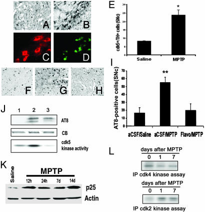
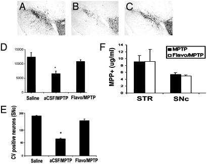
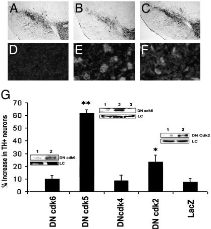
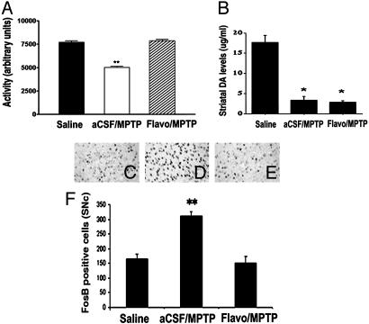
Recent evidence indicates that cyclin-dependent kinases (CDKs, cdks) may be inappropriately activated in several neurodegenerative conditions. Here, we report that cdk5 expression and activity are elevated after administration of 1-methyl-4-phenyl-1,2,3,6-tetrahydropyridine (MPTP), a toxin that damages the nigrostriatal dopaminergic pathway. Supporting the pathogenic significance of the cdk5 alterations are the findings that the general cdk inhibitor, flavopiridol, or expression of dominant-negative cdk5, and to a lesser extent dominant-negative cdk2, attenuates the loss of dopaminergic neurons caused by MPTP. In addition, CDK inhibition strategies attenuate MPTP-induced hypolocomotion and markers of striatal function independent of striatal dopamine. We propose that cdk5 is a key regulator in the degeneration of dopaminergic neurons in Parkinson's disease.


Figures

Fig. 1.
Fig. 1.
Increased cdk5 expression and activity in the SNc of MPTP-treated mice. (A and B) Expression of cdk5 in the SNc of mice treated with saline (A)or MPTP (14 days) (B). (C and D) Representative photomicrographs illustrating the coexpression of cdk5 (D) with TH-positive (C) cells. (E) Quantitative analyses of TH+/cdk5+ neurons. Each bar represents mean ± SEM; * indicates statistical significance (Student's t test, P < 0.01, n = 4). (F-H) Representative photomicrographs showing AT8 expression in the SNc of mice treated with saline (F), MPTP (14 days) and vehicle (G), or MPTP and flavopiridol (H). (I) Quantitative analysis of AT8-positive neurons of the SNc. Each bar represents the mean ± SEM; ** indicates statistical significance (ANOVA, P < 0.001, n = 6-8). (J) Western blot analyses (AT8 expression, 50- to 55-kDa species) and in vitro kinase assay (histone H1 substrate) indicating cdk5 activity in SNc extracts after saline treatment (lane 1), 12 h after chronic MPTP (lane 2), and 14 days after chronic MPTP treatment (lane 3). CB, Coomassie blue stain as loading control. (K) Western blot showing the expression of the cdk5 activator p25 after saline treatment and chronic MPTP treatment at the indicated times. (L) In vitro immunoprecipitate kinase assay showing activity of cdk2 (with histone H1 as substrate) and cdk4 (with Rb as substrate) after MPTP treatment for the indicated times.
Fig. 2.
Fig. 2.
Flavopiridol administration attenuates MPTP-induced degeneration of nigral dopamine neurons. (A-C) Representative TH immunoreactivity in the ventral midbrain of animals treated with saline (A), vehicle/MPTP (B), or flavopiridol/MPTP (C). (D) Quantitative analyses of TH-positive neurons of the SNc. (E) Quantitative analyses of cresyl violet staining at the level of the MTN. Each bar represents the mean ± SEM; * denotes significance (ANOVA, P < 0.0.05, n = 6-8). (F) MPP+ measurements, 90 min after a single-dose MPTP injection into animals treated with either vehicle or flavopiridol (given 24 h before MPTP treatment). Bar graph represents the levels of MPP+ recorded by HPLC analyses (n = 4 or 5 per group) in striatum (STR) and SNc.
Fig. 3.
Fig. 3.
Adenovirus-mediated inhibition of cdk5 provides significant protection from MPTP-induced degeneration of dopaminergic neurons of the SNc. (A-C) Representative photomicrographs of TH immunoreactivity in the ventral midbrain (MTN level) of saline-treated mice (A), mice expressing lacZ and treated with MPTP 14 days earlier (B), and mice expressing DN cdk5 and MPTP (C). Mice were treated with a single unilateral virus injection into the striatum as described in Materials and Methods. (G) Quantitation of TH-positive neurons in the SNc for the indicated treatment groups. Values are described as percent increase in the number of neurons in the ipsilateral (virus-injected) vs. noninjected contralateral control side. Data are represented as mean ± SEM (n = 6 animals per group). * and ** denote significance P < 0.05 and P < 0.001, respectively (ANOVA). (Insets) Expression of DN cdk2, -5, and -6 in SNc extracts by Western blot analyses using an anti-FLAG antibody. (Insets) Lane 1, contralateral control side; lane 2, ipsilateral virus-injected side; and lane 3, noninjected control animal. LC, loading control (Coomassie blue staining). (D-F) Immunofluorescence analyses showing the expression of FLAG-tagged DN proteins in the SNc of animals treated with lacZ (D), DN cdk5 (E), or DN cdk2 (F).
Fig. 4.
Fig. 4.
Flavopiridol administration reduces behavioral impairments and δ FosB (a marker for postsynaptic changes in the denervated striatum levels) after MPTP treatment in mice but does not prevent loss of striatal dopamine. (A) Locomotor activity 14 days after chronic MPTP treatment. Bar graph represents total activity over a 30-min period immediately after amphetamine challenge in a novel open-field test. Each bar represents the mean ± SEM (n = 6-8 animals per group); * indicates statistical significance (P < 0.001, ANOVA). (B) Striatal dopamine content. The striatum was extracted from mice treated with either vehicle/MPTP (14 days) or flavopiridol/MPTP (14 days) and analyzed for dopamine levels by HPLC. Values are expressed as mean ± SEM; * denotes statistical significance (Student's t test, P < 0.01). (C-E) Expression of δ FosB in the striatum of mice treated with saline (C), vehicle and MPTP (14 days) (D), or flavopiridol and MPTP (14 days) (E). (F) Quantitation of δ FosB-positive cells; each bar represents mean ± SEM; * indicates statistical significance (P < 0.05, ANOVA, n = 6 animals per group).

References

    1. Przedborski, S., Jackson-Lewis, V., Naini, A. B., Jakowec, M., Petzinger, G., Miller, R. & Akram, M. (2001) J. Neurochem. 76, 1265-1274. - PubMed
    1. Pines, J. (1993) Biochem. Soc. Trans. 21, 921-925. - PubMed
    1. Freeman, R. S., Estus, S. & Johnson, E. M., Jr. (1994) Neuron 12, 343-355. - PubMed
    1. Gao, C. Y. & Zelenka, P. S. (1995) Exp. Cell Res. 219, 612-618. - PubMed
    1. O'Hare, M. J., Hou, S. T., Morris, E. J., Cregan, S. P., Xu, Q., Slack, R. S. & Park, D. S. (2000) J. Biol. Chem. 275, 25358-25364. - PubMed

Publication types

MeSH terms