. 2003 Nov 11;100(23):13501-6.
doi: 10.1073/pnas.2135380100. Epub 2003 Oct 31.

Pyrin binds the PSTPIP1/CD2BP1 protein, defining familial Mediterranean fever and PAPA syndrome as disorders in the same pathway

Affiliations

Pyrin binds the PSTPIP1/CD2BP1 protein, defining familial Mediterranean fever and PAPA syndrome as disorders in the same pathway

Nitza G Shoham et al. Proc Natl Acad Sci U S A. .

Abstract

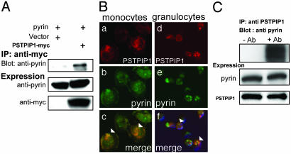
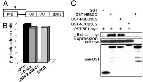
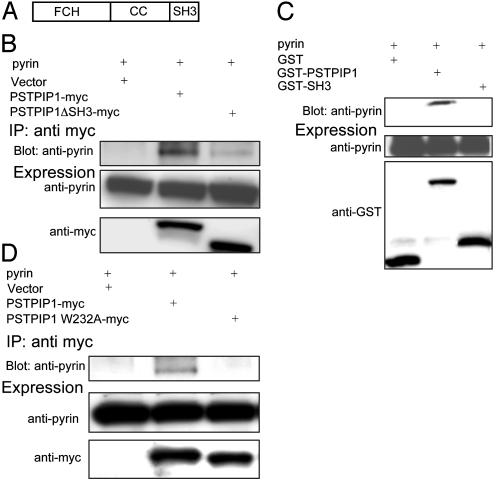
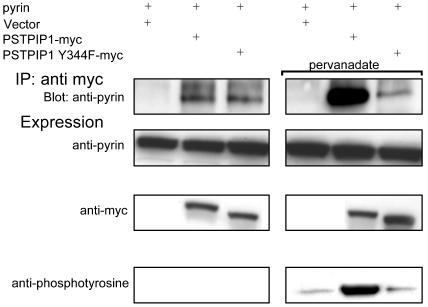
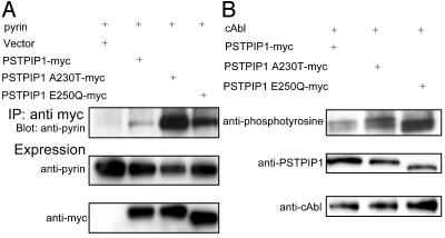
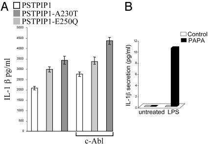
Pyrin, the familial Mediterranean fever protein, is found in association with the cytoskeleton in myeloid/monocytic cells and modulates IL-1beta processing, NF-kappaB activation, and apoptosis. These effects are mediated in part through cognate interactions with the adaptor protein ASC, which shares an N-terminal motif with pyrin. We sought additional upstream regulators of inflammation by using pyrin as the bait in yeast two-hybrid assays. We now show that proline serine threonine phosphatase-interacting protein [PSTPIP1, or CD2-binding protein 1 (CD2BP1)], a tyrosine-phosphorylated protein involved in cytoskeletal organization, also interacts with pyrin. Recently, PSTPIP1/CD2BP1 mutations were shown to cause the syndrome of pyogenic arthritis, pyoderma gangrenosum, and acne (PAPA), a dominantly inherited autoinflammatory disorder mediated predominantly by granulocytes. Endogenous PSTPIP1/CD2BP1 and pyrin are coexpressed in monocytes and granulocytes and can be coimmunoprecipitated from THP-1 cells. The B box segment of pyrin was necessary and the B box/coiled-coil segment sufficient for this interaction, whereas the SH3 and coiled-coil domains of PSTPIP1/CD2BP1 were both necessary, but neither was sufficient, for pyrin binding. The Y344F PSTPIP1/CD2BP1 mutation, which blocks tyrosine phosphorylation, was associated with a marked reduction in pyrin binding in pervanadate-treated cells. PAPA-associated A230T and E250Q PSTPIP1/CD2BP1 mutations markedly increased pyrin binding as assayed by immunoprecipitation and, relative to WT, these mutants were hyperphosphorylated when coexpressed with c-Abl kinase. Consistent with the hypothesis that these mutations exert a dominant-negative effect on the previously reported activity of pyrin, we found increased IL-1beta production by peripheral blood leukocytes from a clinically active PAPA patient with the A230T PSTPIP1/CD2BP1 mutation and in cell lines transfected with both PAPA-associated mutants.


Figures

Fig. 1.
Fig. 1.
Pyrin interacts with PSTPIP1. HeLa cells were transiently cotransfected with PSTPIP1-myc (or vector) and pyrin-expressing plasmids. (A) Lysates were incubated with anti-myc agarose beads and washed, and eluates were separated on SDS/PAGE. Blots were probed with rabbit polyclonal antipyrin and with mouse monoclonal anti-myc. (B) Adherent human monocytes (a-c) and human granulocytes (d-f) were fixed and immunostained with polyclonal rabbit anti-PSTPIP1 followed by Alexa Fluor-568-conjugated goat anti-rabbit IgG to detect endogenous PSTPIP1 (a and d, red) and with Alexa Fluor-488-conjugated rabbit anti-pyrin (b and e, green). (c and f) Merge of PSTPIP1 and pyrin cellular localization images) demonstrating colocalization. Arrowheads indicate regions where staining overlap was most significant. (C) To assess endogenous protein interactions, lysates of THP-1 cells were subjected to immunoprecipitation with crosslinked rabbit polyclonal anti-PSTPIP1 (+Ab) or control serum (-Ab). Lysates were washed and separated on SDS/PAGE. Blots were probed with rabbit polyclonal anti-pyrin or anti-PSTPIP1.
Fig. 2.
Fig. 2.
The B box domain of pyrin is necessary for the interaction with PSTPIP1. (A) Schematic diagram of the structural domains of pyrin. PYD, PYRIN domain; BB, B box; CC, coiled-coil. “N” denotes amino acids 1-374 (not to scale). (B) Yeast cells were cotransformed with WT pyrin or mutant constructs in pDBLeu and with PSTPIP1 in pPC86. Cells were subjected to a quantitative liquid culture assay for β-galactosidase activity. β-Galactosidase units indicate absorbance at 574 nm, normalized to culture density and incubation time, and represent the degree of interaction between PSTPIP1 and various pyrin mutants. (C) Cos 7L cells were cotransfected with PSTPIP1-myc and GST, GSTNBBCC, GST-NBBB30.2, or GST-NCCB30.2. GST-NBBCC was transfected without PSTPIP1-myc, as another control. Lysates were precipitated with glutathioneagarose beads, washed, eluted, and immunoblotted with anti-myc. Protein expression levels were confirmed with anti-GST or anti-myc.
Fig. 3.
Fig. 3.
The SH3 and coiled-coil domains of PSTPIP1 are necessary, but neither is sufficient for pyrin binding. (A) Schematic diagram of the structural domains of PSTPIP1. FCH, Fer-CIP4 homology domain; CC, coiled-coil (not to scale). (B) HeLa cells were transiently cotransfected with pyrin and with PSTPIP1-myc, PSTPIP1ΔSH3-myc, or vector. Lysates were immunoprecipitated with anti-myc and immunoblotted with anti-pyrin and with anti-myc. (C) HeLa cells were cotransfected with pyrin and GST, GST-PSTPIP1, or GST-SH3. Lysates were precipitated with glutathione-agarose beads, washed, eluted, and immunoblotted with anti-pyrin. Protein expression levels were confirmed with anti-GST or anti-pyrin. (D) HeLa cells were cotransfected with pyrin and myc-tagged PSTPIP1, PSTPIP1 W232A, or vector. Lysates were immunoprecipitated with anti-myc and immunoblotted with anti-pyrin or anti-myc.
Fig. 4.
Fig. 4.
Tyrosine phosphorylation of PSTPIP1 increases PSTPIP1-pyrin binding. HeLa cells were transiently cotransfected with pyrin and with PSTPIP1-myc, PSTPIP1 Y344F-myc, or vector. Cells were either untreated (Left) or were incubated for 10 min with the phosphatase inhibitor pervanadate before immunoprecipitation (Right). Lysates were immunoprecipitated with anti-myc and immunoblotted with anti-pyrin, anti-myc, or antiphosphotyrosine. Phosphorylation of PSTPIP1 was examined with antiphosphotyrosine (Bottom).
Fig. 5.
Fig. 5.
PAPA-associated PSTPIP1 mutations increase PSTPIP1-pyrin binding and are hyperphosphorylated. (A) HeLa cells were transiently cotransfected with pyrin and PSTPIP1-myc, PSTPIP1 A230T-myc, PSTPIP1 E250Q-myc, or vector. Lysates were immunoprecipitated with anti-myc and immunoblotted with anti-pyrin and anti-myc. (B) 293T cells were transiently cotransfected with WT PSTPIP1-myc or myc-tagged mutants and cAbl. Proteins were immunoprecipitated with anti-myc agarose beads. Tyrosine phosphorylation of WT PSTPIP1 or PSTPIP1 A230T and PSTPIP1 E250Q was detected by blotting with antiphosphotyrosine (Top). Protein expression levels were confirmed by immunoblot with anti-PSTPIP1 (Middle) or anti-cAbl (Bottom).
Fig. 6.
Fig. 6.
Effect of PAPA-associated PSTPIP1 mutations on IL-1 secretion. (A) COS-7L cells were cotransfected with expression plasmids for mouse IL-1β and human pyrin, ASC, caspase-1, and PSTPIP1, with or without c-Abl. Supernatants were assayed for IL-1β at 24 h (mean of four replicate experiments). Without c-Abl, WT vs. A230T, P < 0.01, WT vs. E250Q, P < 0.003; with c-Abl, WT vs. A230T, P < 0.004, WT vs. E250Q, P < 0.008. (B) Adherent monocytes from a patient with active PAPA syndrome or healthy controls either were not treated or were stimulated with 1 μg/ml lipopolysaccharide for 1 h. Supernatants were assayed for IL-1β.

References

    1. Galon, J., Aksentijevich, I., McDermott, M. F., O'Shea, J. J. & Kastner, D. L. (2000) Curr. Opin. Immunol. 12, 479-486. - PubMed
    1. Kastner, D. L. (2003) in Rheumatology, eds. Hochberg, M. C., Silman, A. J., Smolen J. S., Weinblatt, M. E. & Weisman, M. H. (Mosby, Edinburgh), 3rd Ed., pp. 1717-1734.
    1. International FMF Consortium (1997) Cell 90, 797-807. - PubMed
    1. French FMF Consortium (1997) Nat. Genet. 17, 25-31. - PubMed
    1. Centola, M., Wood, G., Frucht, D. M., Galon, J., Aringer, M., Farrell, C., Kingma, D. W., Horwitz, M. E., Mansfield, E., Holland, S. M., et al. (2000) Blood 95, 3223-3231. - PubMed

MeSH terms